Olympic B3 Science Summer Camp 2014
Biotechnology, Biodiversity and Bioinformatics
June 11th - June 26th
Pictures of Individuals and Class Activities were posted daily -- Also a Blog by Annabel Richards
Facebook page has even more pictures but fewer captions and can be found at this link Requires Login
June 11 th
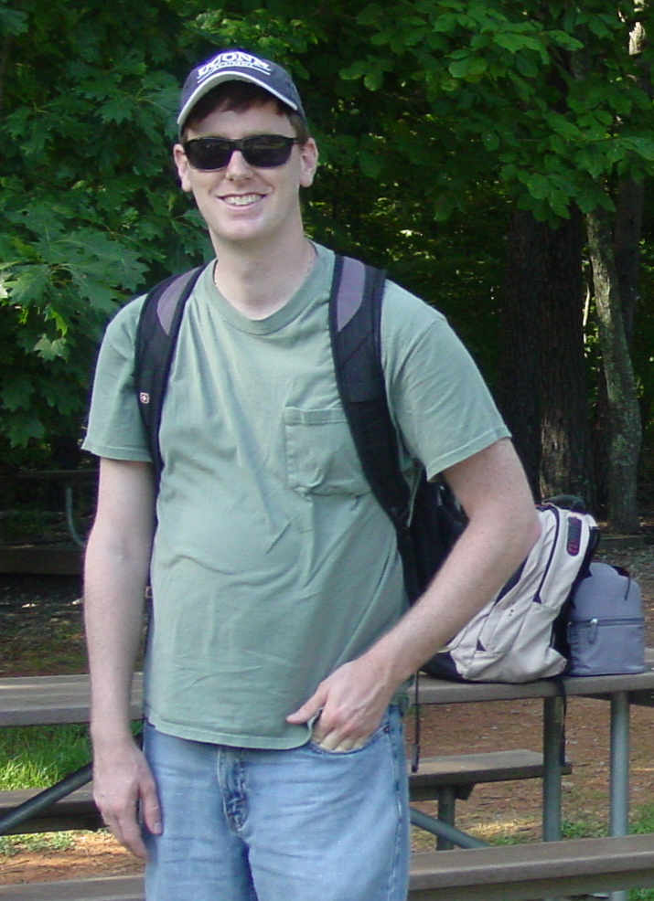
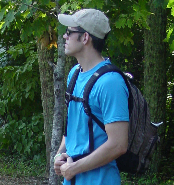
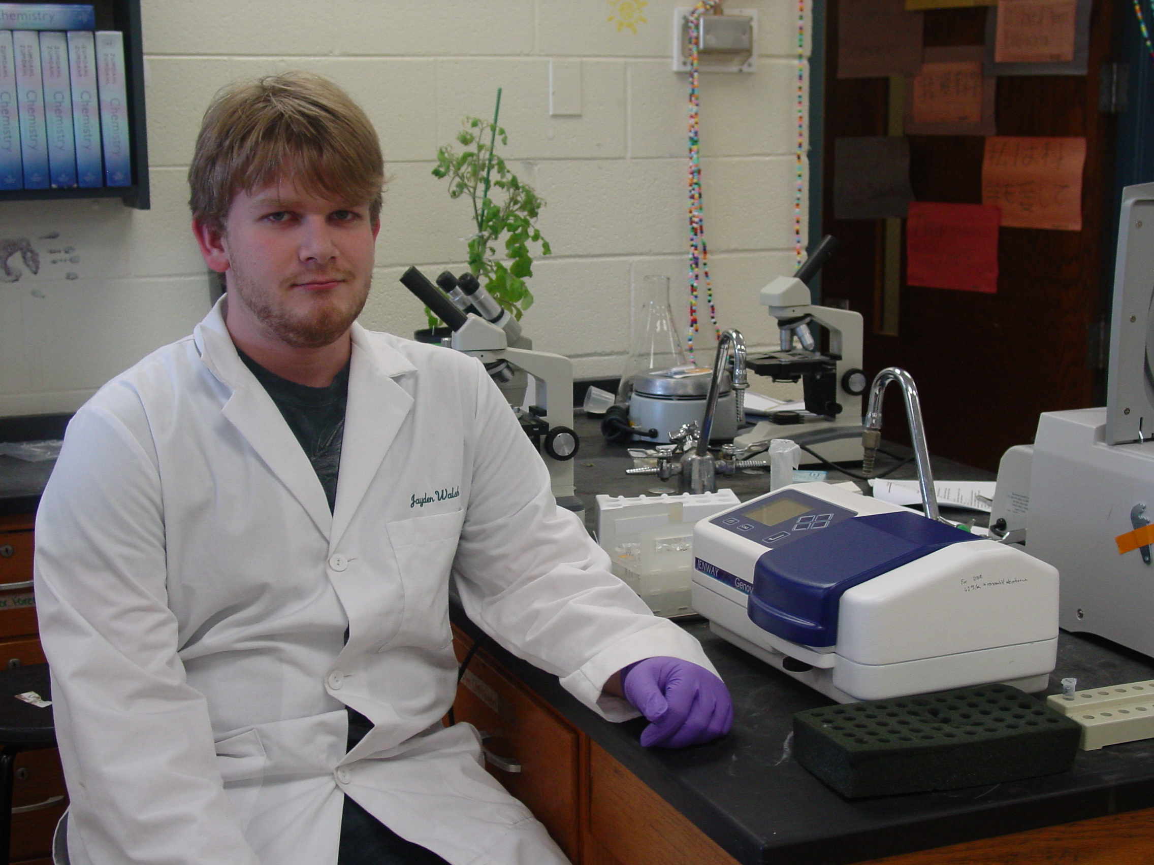
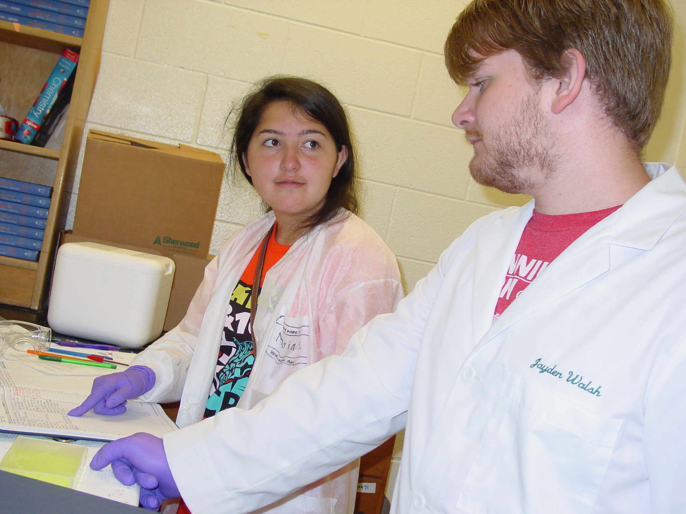
Instructors and TA
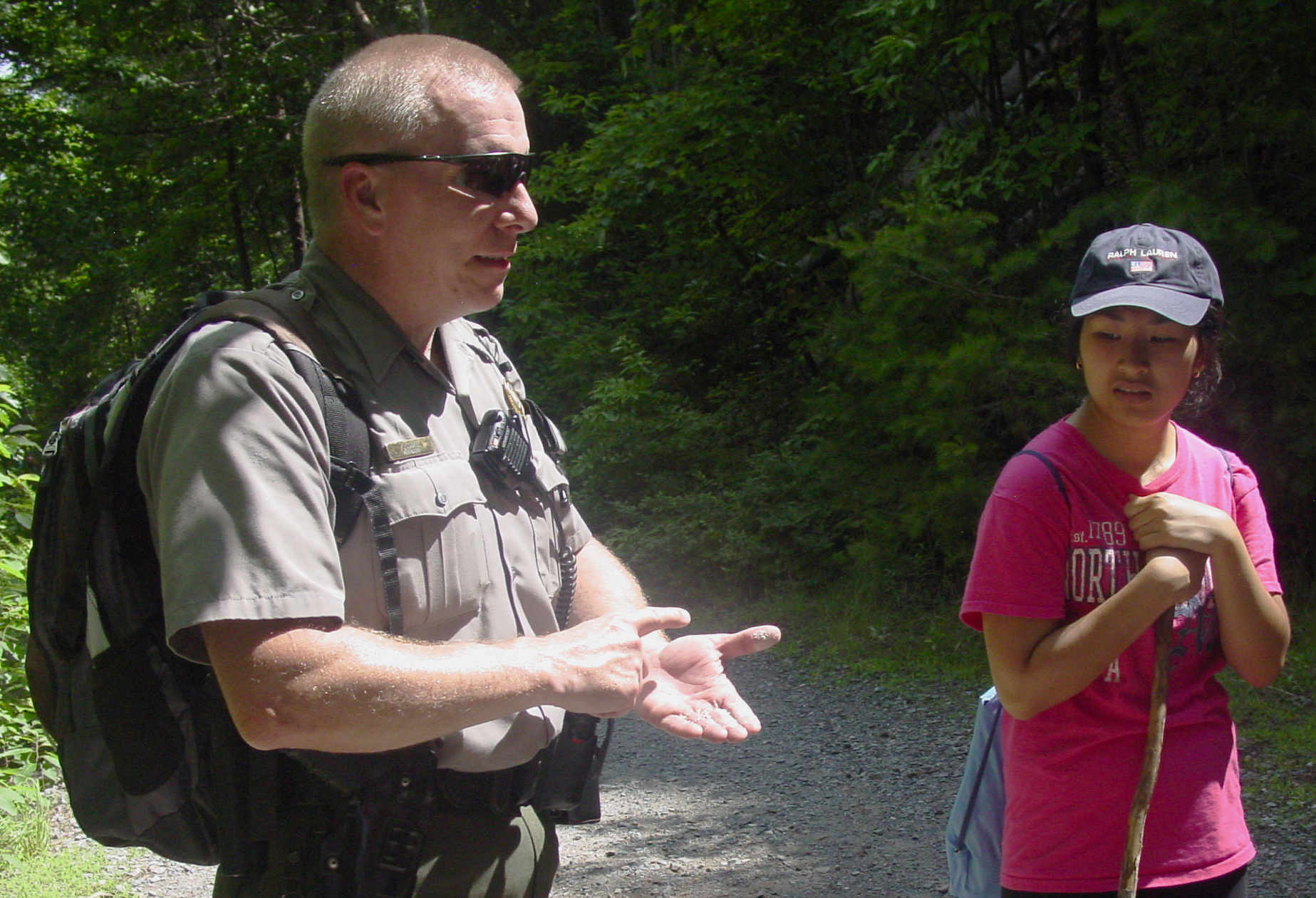
Ms Smith. Jayden Walsh (TA), Dr. Weller, Ms Putnam and Ms Smith and students. Guests: Steve Barilovits of The American Chestnut Foundation and Ranger Kelly Cook of the NC State Parks.
Steve and Kelly. Steve Lecturing .Kelly Lecturing. Annabel Richards, our class journalist. Getting the Lab ready: Meilee and Jayden worked hard to get the lab set up (with help from others).
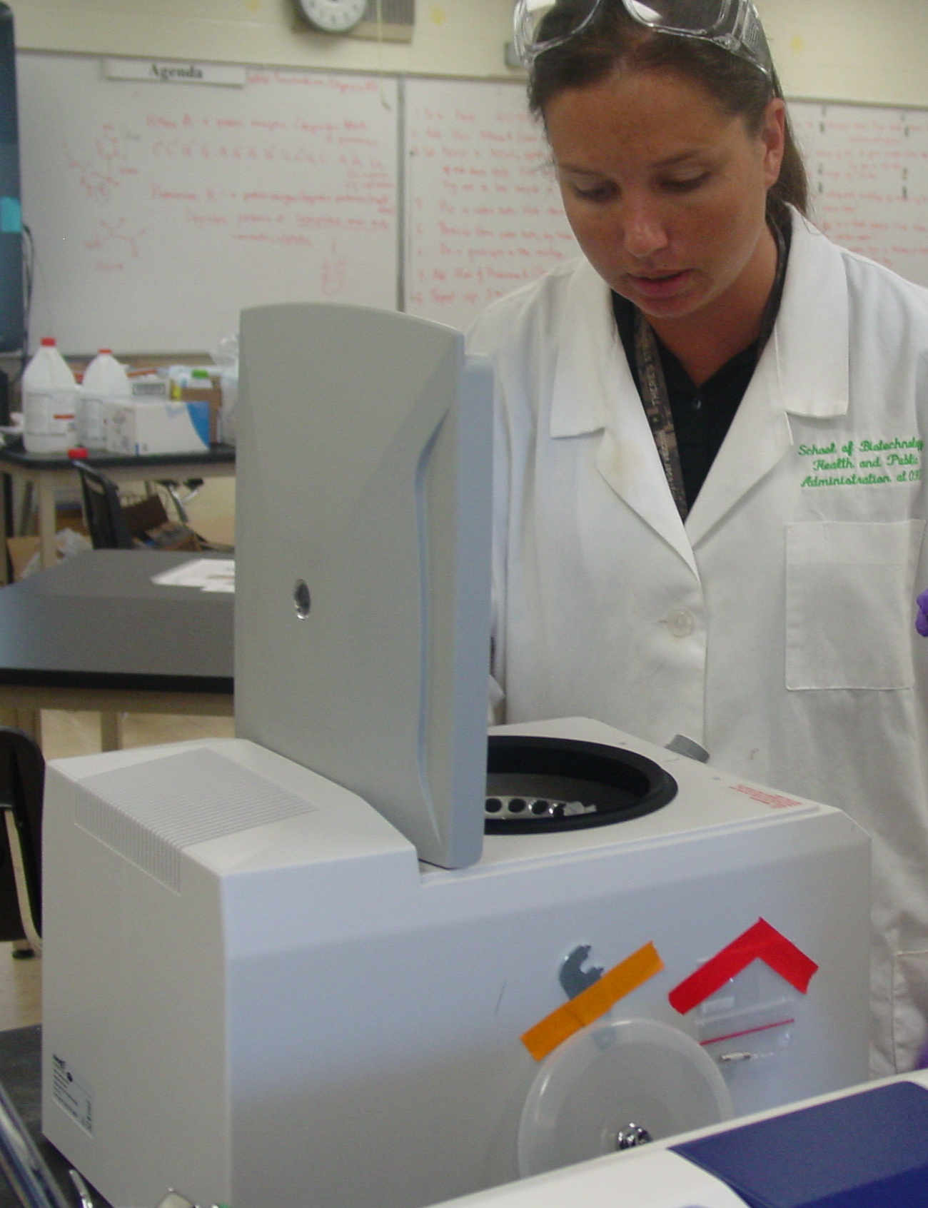
Before they started Meilee putting down bench paper Sorting out the materials We had students test their knowledge of our most frequently-used pieces of equipment.
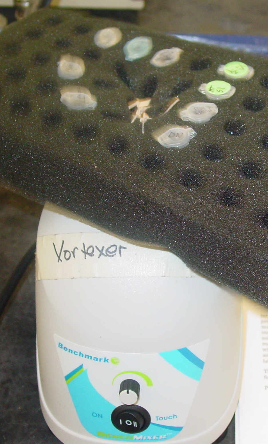
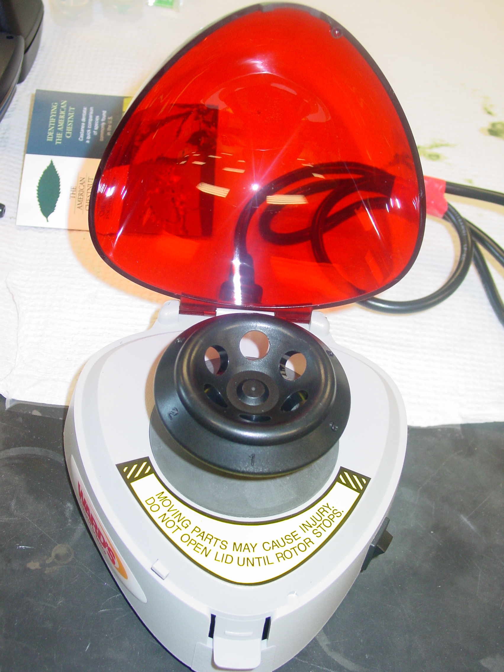
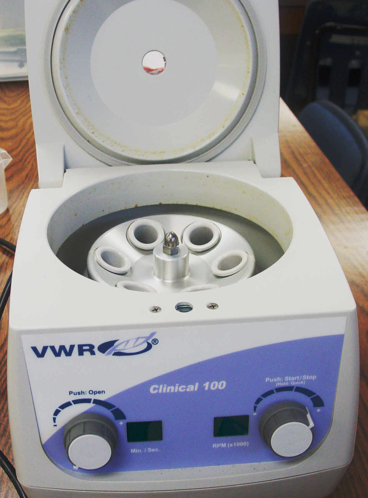
Micropipettes PCR machine Electrophoresis Rig Minifuge Spectrophotometer Waterbath Vortexer In preparation for our field trips, students examined leaves and pollen with eyes and microscopes and recorded observations in their notebooks.
Hoa checking out leaves Doudgie at the 'scope. Maria making art of a notebook Tripp, Nicole, Tamden and Sanjana. Steve and Meilee examining Chestnut flowers Meilee recording the data in her notebook. Many students were not captured in action today, it was a bit chaotic. They will appear on future dates.
Our class journalist, Annabel Richards, is contributing a daily blog, to show the students view of our activities.
Wednesday, June 11 th, 2014. The B3 Summer Science Camp Blog
What an energetic atmosphere I observed today! It is clear that many of the students involved
in this program are eager to begin making hands-on discoveries and obtaining knowledge for themselves. Today, we received the basic
"throw-down" of what we are trying to accomplish, how we are going to accomplish this, and the importance of our effort. To sum this up,
we are trying to save the American Chestnut tree from the Chestnut blight fungus, Cryphonectria parasitica. This particular fungus began
to claim the lives of MANY American Chestnut trees around the early 1900s. Today, we are still searching for a way to cross-breed these
living organisms and discover a way to make them resistant to the blight. We can do this by discovering one or more genes in the Chestnut
genome that allow the American Chestnut tree to slowly evolve immunity over generations. We may not be around to witness the achievement
of this, because it takes many years, but our efforts will mean a world of difference to scientists in the future with similar ambitions.
I am honored to witness so many individuals take part in such a unique opportunity. In such a busy world, it is beautiful for a group of
individuals to come together, and devise a plan to give something back to our planet. Tomorrow we are going to take our first field trip
over to the American Chestnut Foundation orchard. Hopefully the weather is lovely! ----- Annabel Richards
June 12 th
Field Trip at Pryor's Farm in Hendersonville, NC
We arrived at the Pryor Farm, which has an experimental plantation of American Chestnut crosses at 10am. The goals were to: bag chestnut flowers, inoculate young trees with fungus to test their resistance, and weed.
View from the Pryor Farm The B3 class ready to go to work. The Pryor Farm Plantation of young trees to be tested for blight resistance. Dr. Paul Sisco, the scientist guiding our days work.
We joined a high school group from Brevard, to maximize our effect
The Brevard group counting off to make teams. Part I: Weeding!
Hand weeding means chemicals like RoundUp can be avoided: chemicals will kill young trees.
Rows being cleared of weeds. James, Nicole, Sanjana and Tamden, ace weeding team.
Dirt and unexpected bugs happened.
Joint Olympic-Brevard dirt patrol Ms Putnam finds ???? Willie also found Something Part II: Bagging Female Flowers
Bagging female flowers means you can control their fertilization, and get the cross you want to test in the next generation.
First, the tree must be flowering.
A flowering tree behind Dr. Sisco. Showing the reproductive parts.
The female flower must be at the right stage: mature but not completely open.
Female flowers (frilly and light green) surrounded by male flowers (pebbly and yellow in long strands).
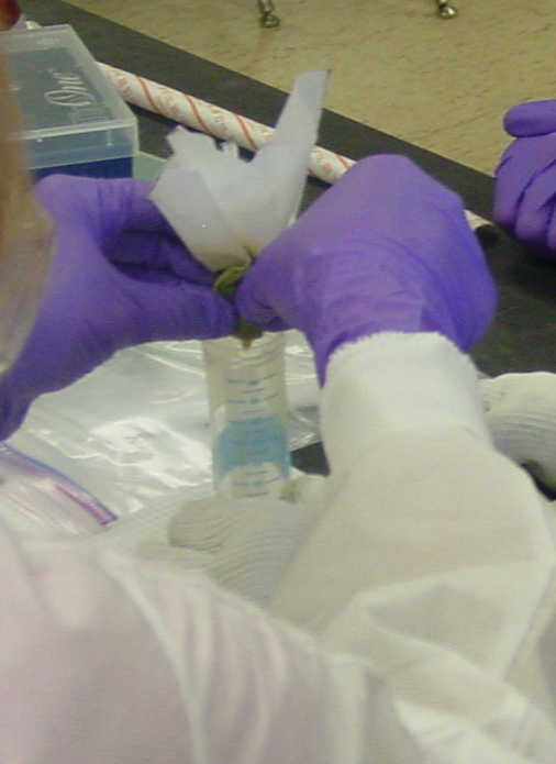
You have to strip away the nearby male flowers and leaves, back 5-6 inches on the stem, then bag the flower to protect it from random pollen.
Cutting back male flowers with scissors. Ms Smith Demonstrates. Doudgie and partner placing a bag Part III: Inoculating young trees with fungus to see if they are resistant
This was the most complicated technique we learned. A small hole is made in the bark, a plug containing the fungus is inserted and tape seals it up.
First, a young tree is identified and the bark is sterilized by wiping it with alcohol.The a small hole is made through the bark.
A young tree. Sterilizing the bark. Making a hole in the bark
Fungus has been grown on an agar plate, and round punches are made on the outside rim of the growth area to prepare plugs.
An agar plate of fungus. Using a punch to make plugs in the agar.
You need to flame-sterilize the spatula before you lift out plugs and inoculate the trees. Alcohol is used, and good technique is important so you don't burn yourself.
The sterilization technique. Removing the plug.
After appying the agar plug to the hole in the bark, you seal it over with masking tape so rain won't wash it out. Applying the plug with the spatula. Taping over the infection site. Part IV: Lunch and learn
It started to rain just as we were finished, so we retreated to the Justus Western Justice Academy cafeteria for lunch.
Dr. Sisco eplained the genetics of disease resistance in these chestnut trees.
Explaining multiple alleles. Question and Answer time.
Thanks to a number of people who came along to assist us:
Three scientists from UNC Charlotte Bioinformatics and Genomics: Dr. Cathy Moore, Jon Shea, and Matthew Brown. Tegan Love came as a community volunteer. Thursday, June 12 th, 2014. The B3 Summer Science Camp Blog
Today we took out first field trip up to one of the orchards that belong to the American Chestnut Foundation near Asheville, North Carolina. Needless to say, the grass was extremely itchy
and we were operating under the threat of thunderstorms. The fun, however, came from what we were doing. Those in the B-3 program had the unique opportunity to actually infect a good number of
the cross-bred American chestnut trees with the fungal blight. They were also able to learn about what it meant to bag the trees, and, best of all, they were able to weed! Students had the
opportunity to learn about the unique programs and techniques that TACF is following. I, for one, found the program dealing with the genetically-modified American chestnut trees most fascinating.
When the fungal blight attacks the American chestnut tree, it releases a substance called oxalic acid, which basically kills what it touches so the fungus can come along and eat those cells right up.
The genetically-modified American Chestnut tree, however, contains a special code for oxalic acid oxidase (taken from wheat), which degrades that acid. When oxalic acid and the oxidase come into contact with one another,
neutralization occurs and the acid can no longer harm the tree. Pretty cool, huh?
----- Annabel Richards
June 13 th
Field Trip to Crowder's Mountain State Park to collect samples for analysis
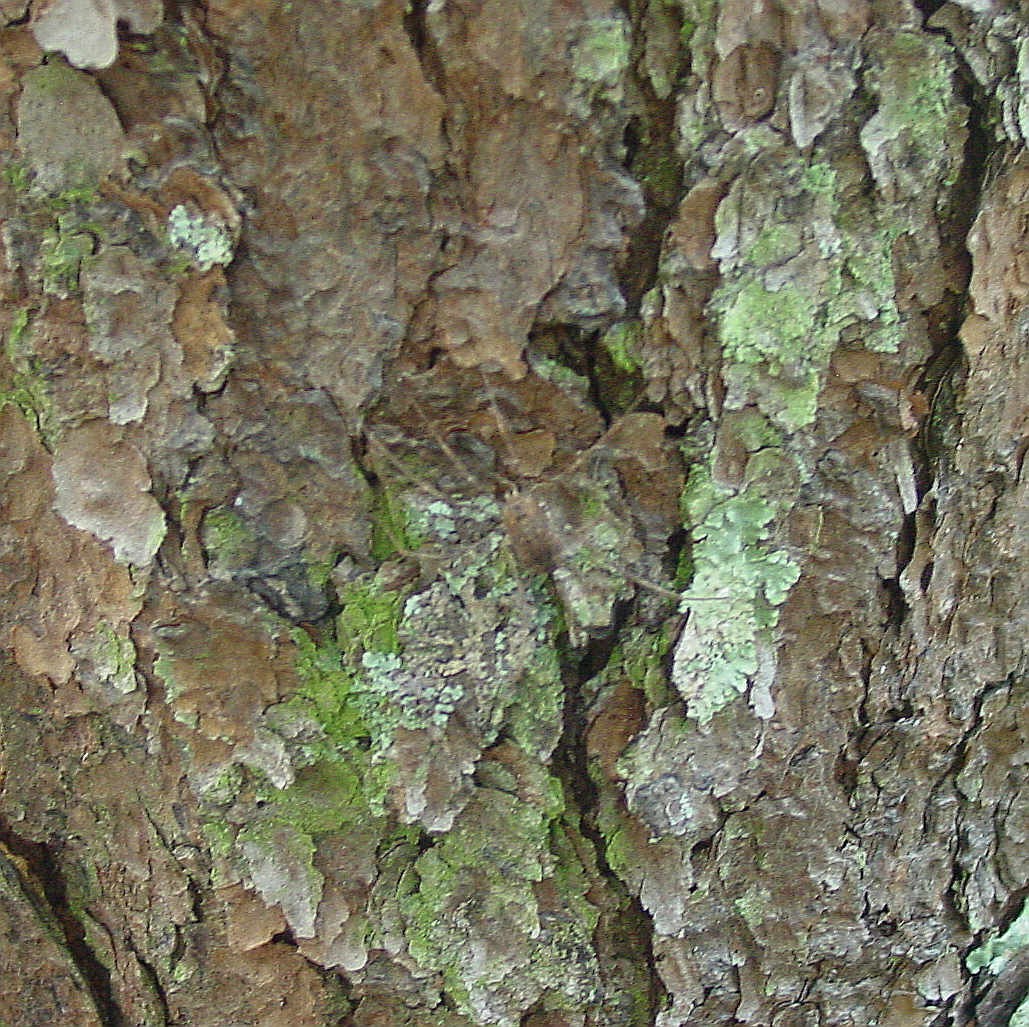
The goal of this field trip was to see the native habitat of a few remaining wild American Chestnuts and to collect some of their leaves.
All have been affected by the blight, so we also hoped to collect some of the fungus.
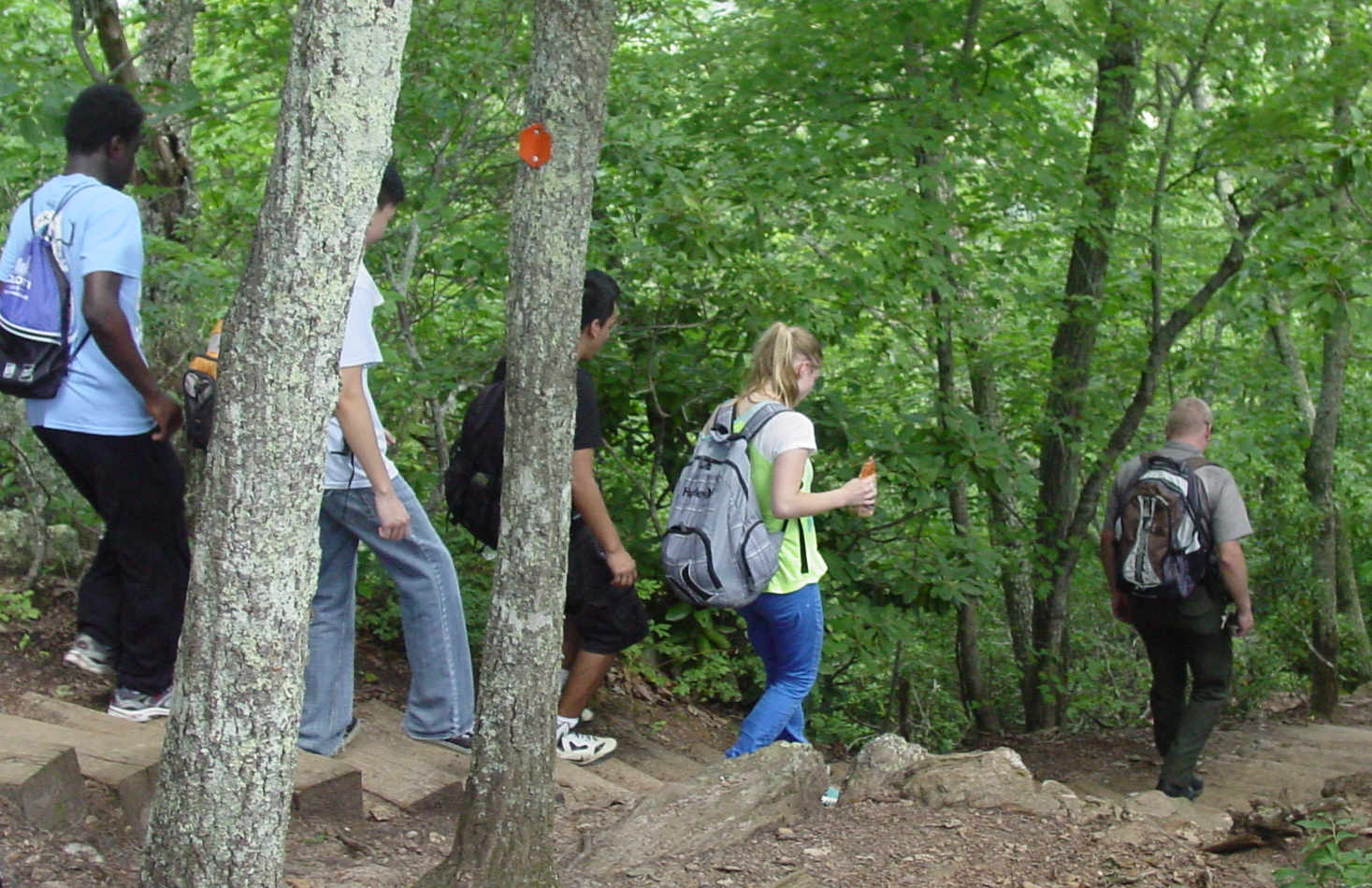
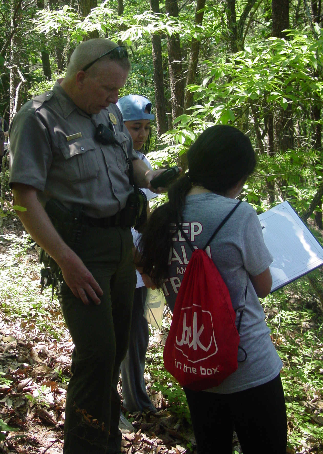
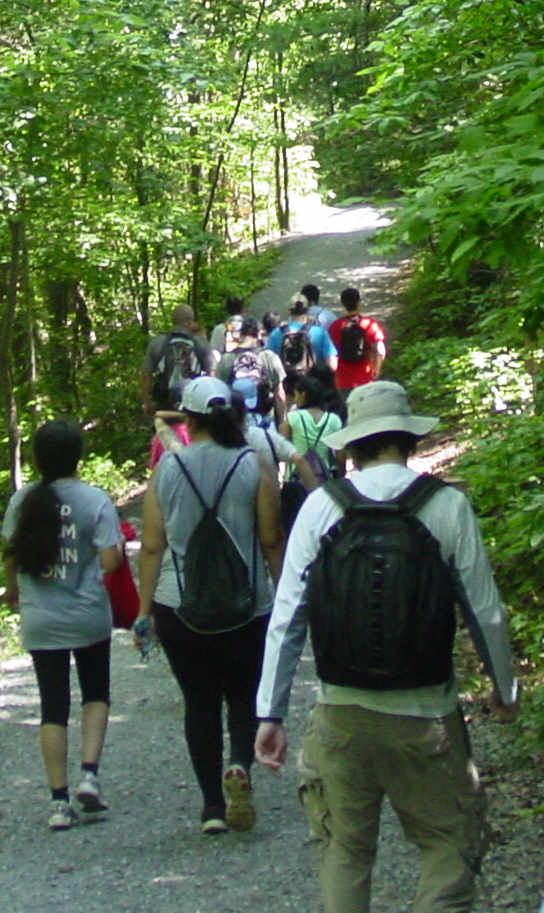
Ranger Kelly Cook was our guide for this trip, which started at the Linwood parking lot and took us to the peak of Crowder's Mountain.
We continued on to an overlook and then looped around the back trail, collecting at several sites along the way.
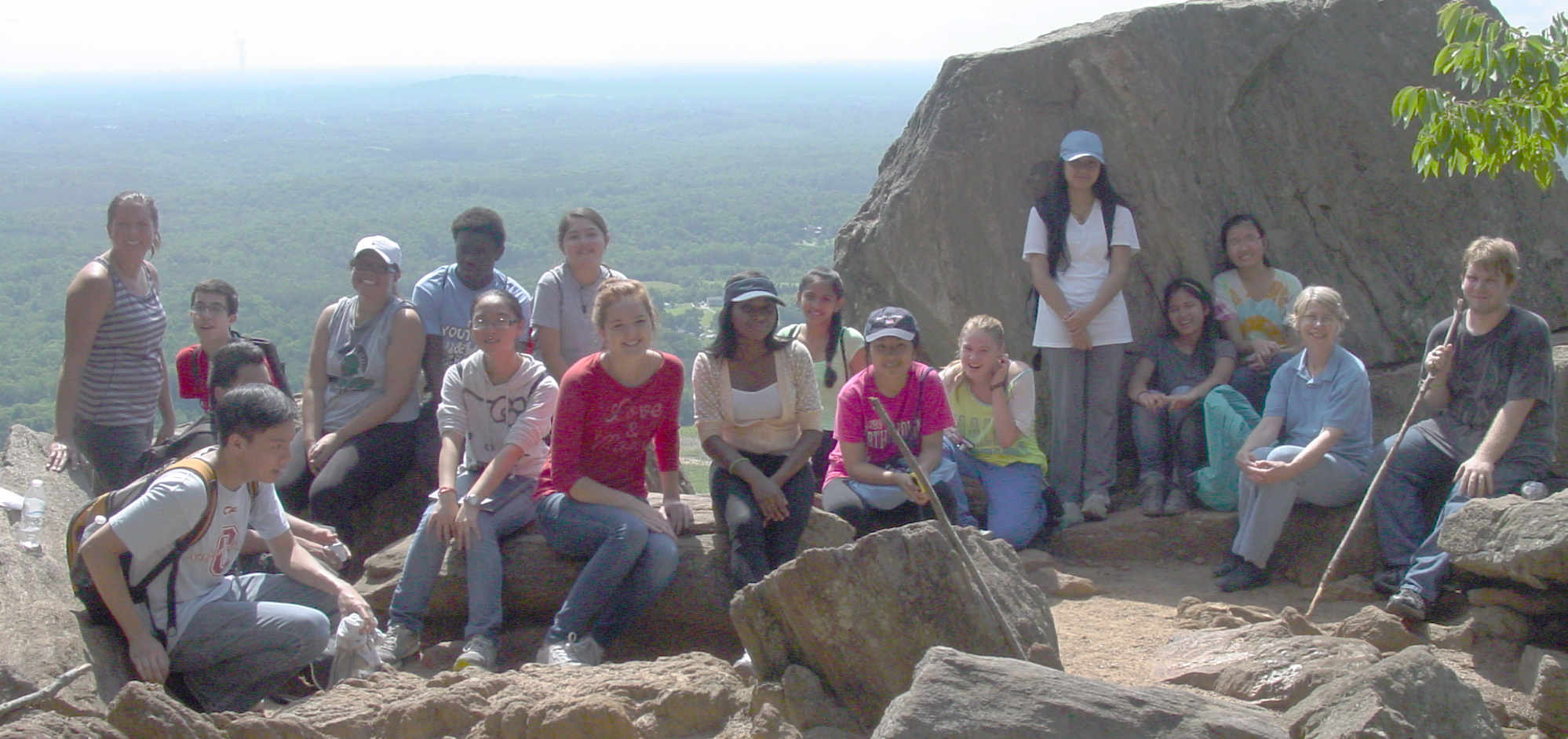
Group pictures start us out, the first at the Linwood parking lot and the second at the scenic overlook near the top of Crowder's Mountain.
We were accompanied again by 3 UNC Charlotte scientists.
Matthew Brown Jon Shea Timm Hamp
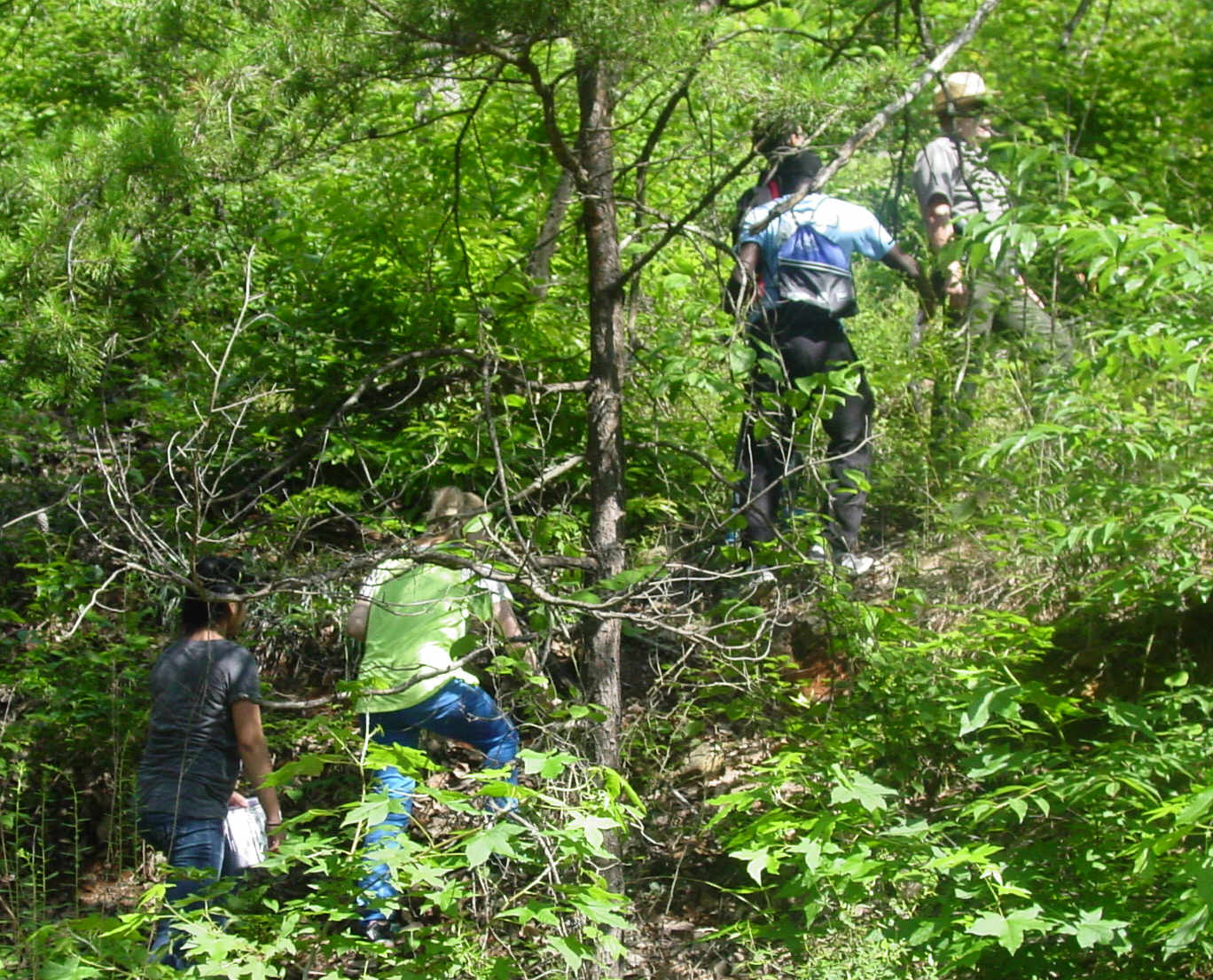
Our first tree was on the Tower Trail, about 1.8 miles up. The approach required some uphill scrambling
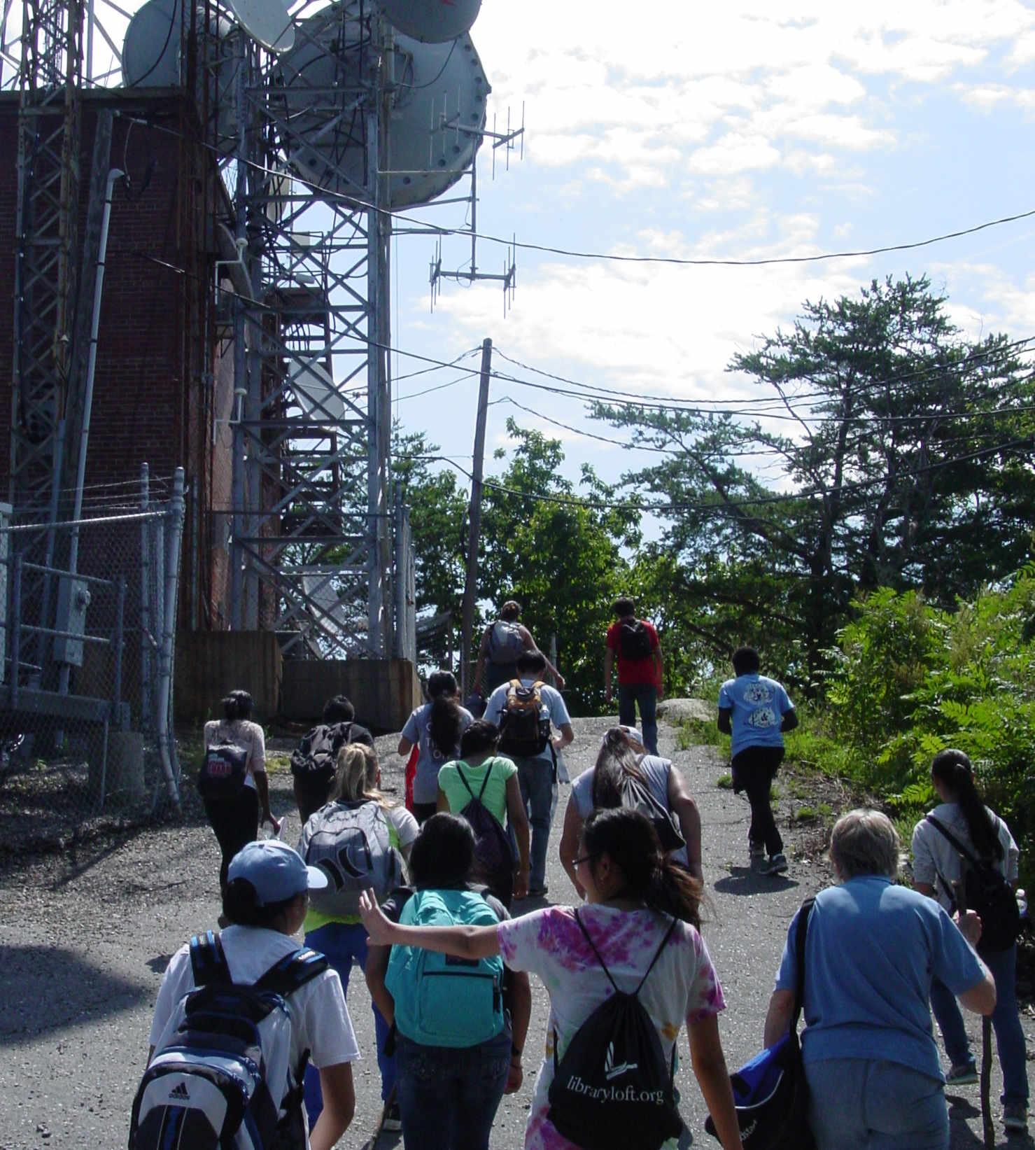
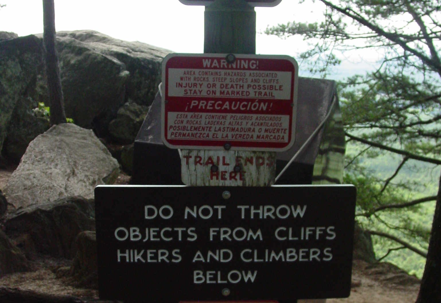
We continued up to the top of Crowder's Mountain, which showcases some very scenic cell phone and TV dishes.
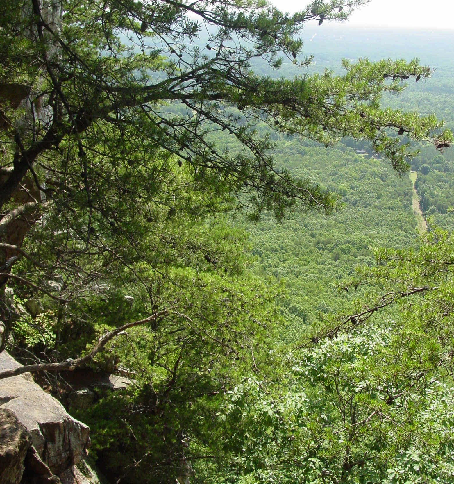
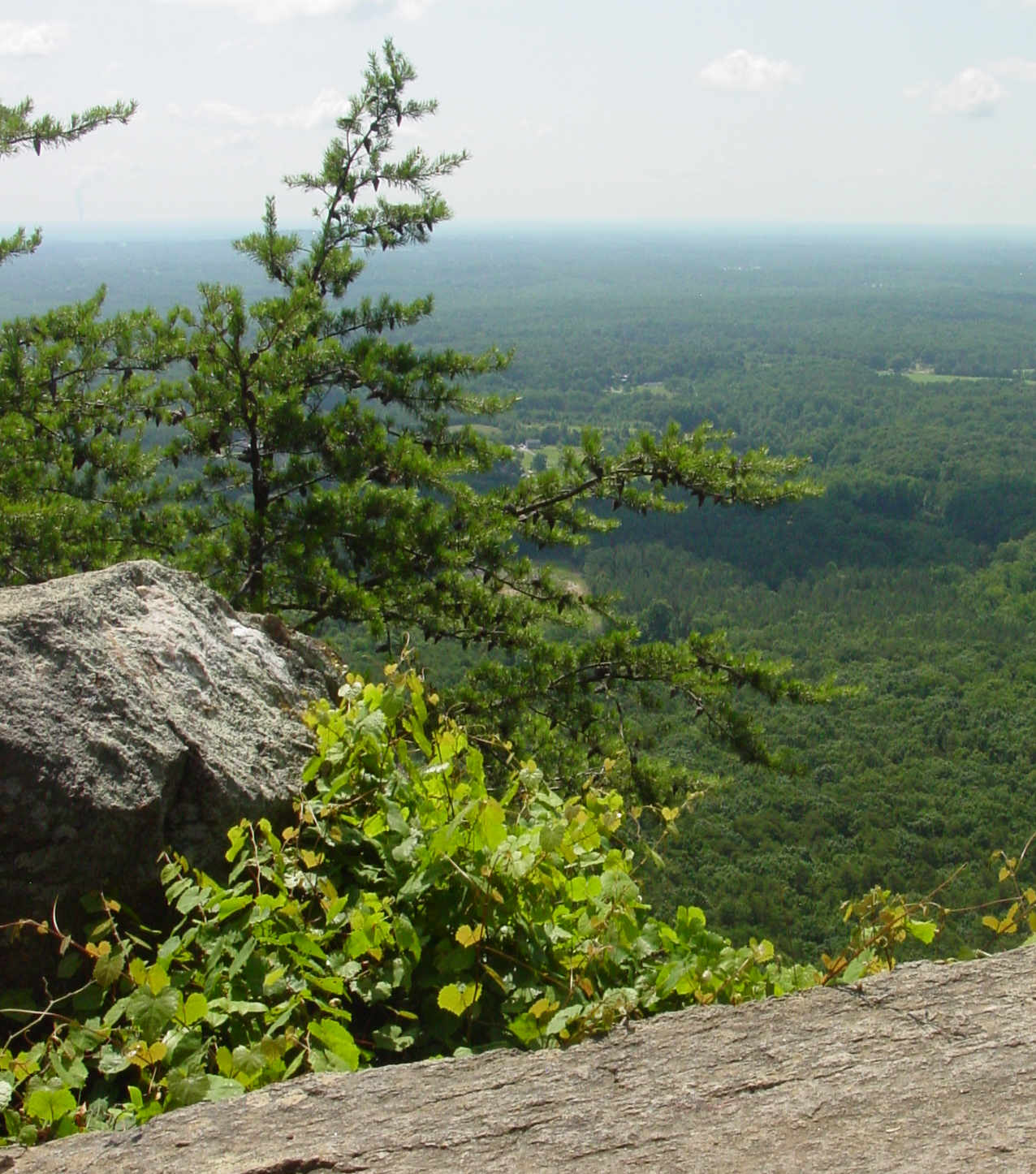
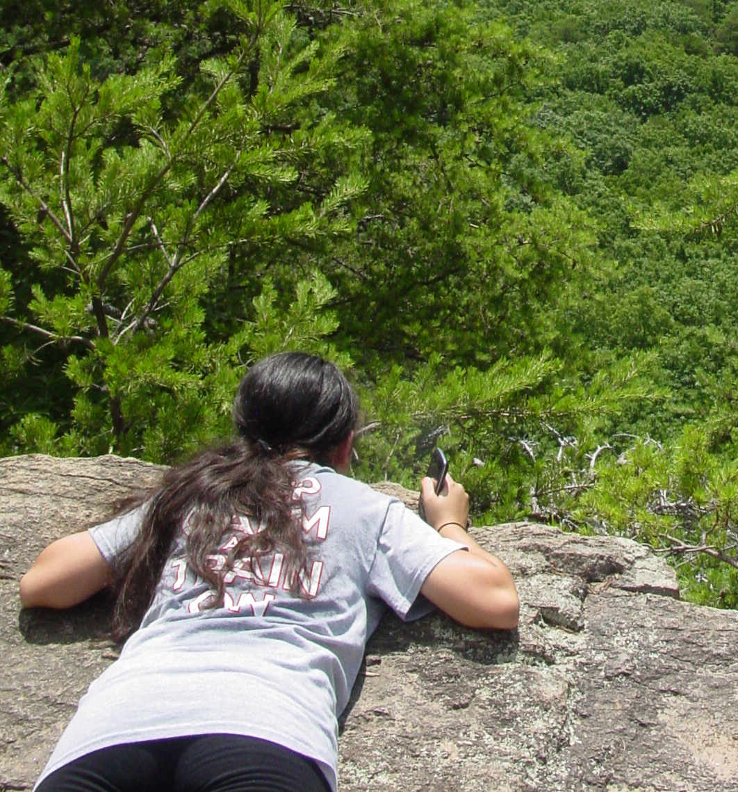
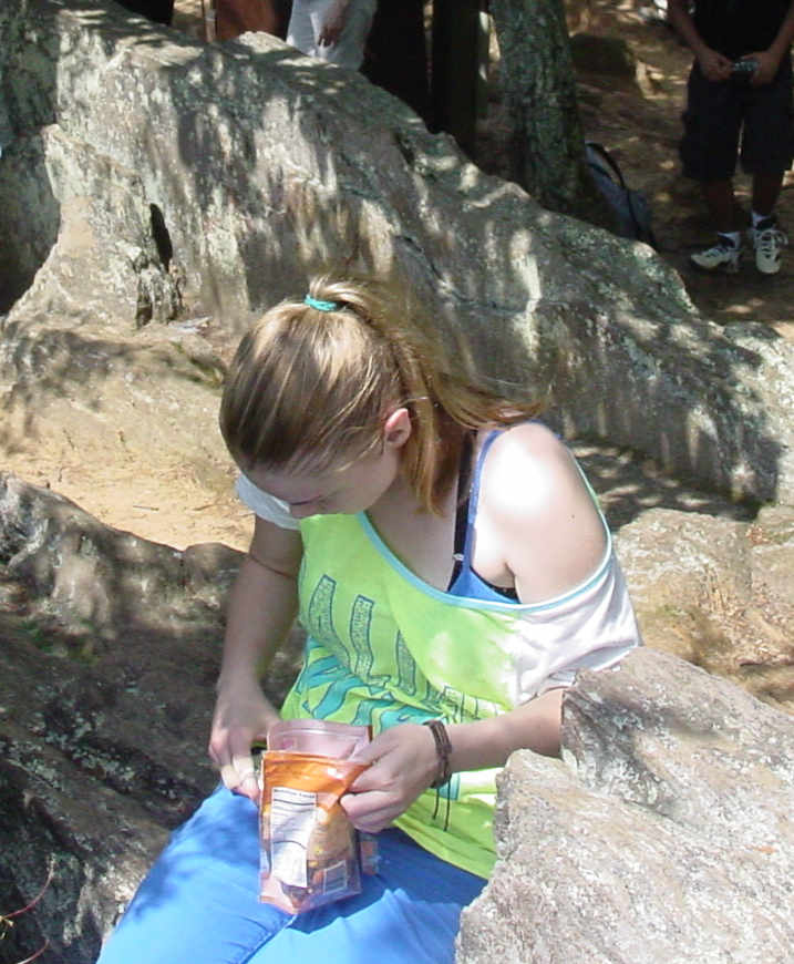
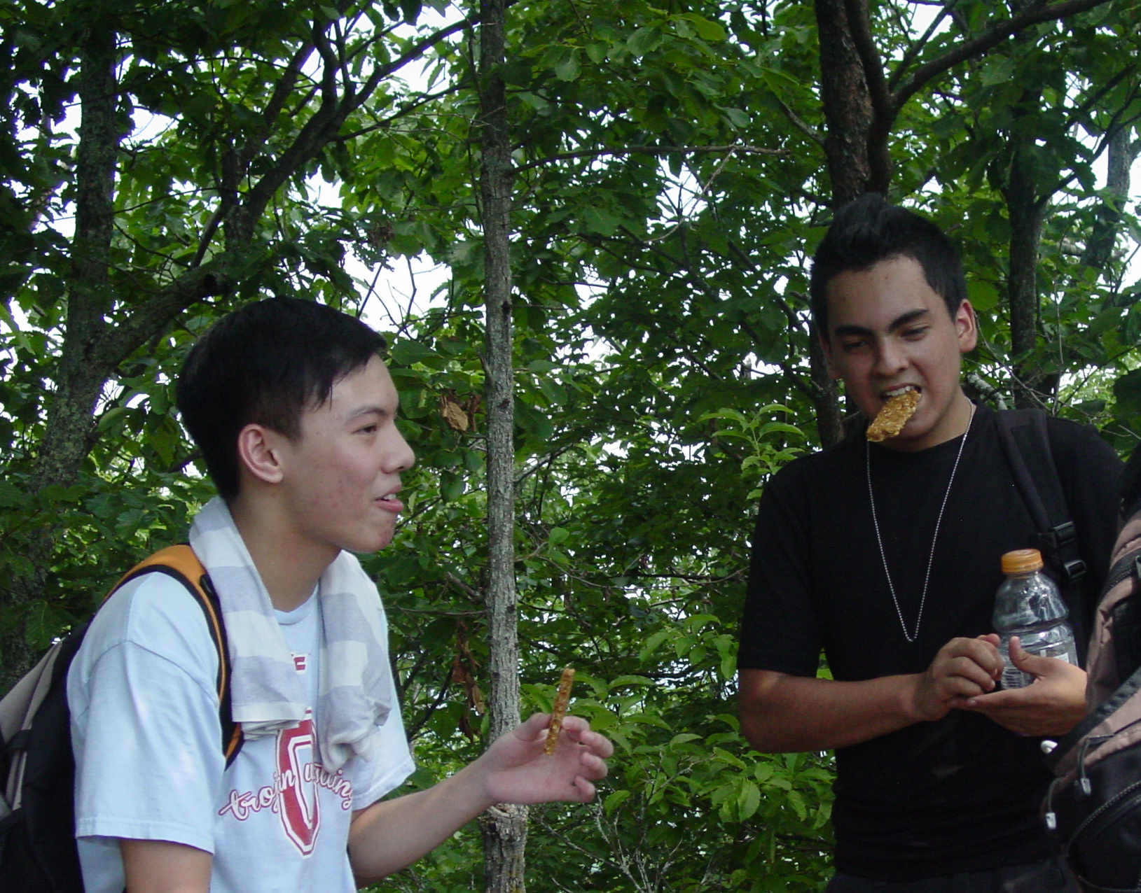
From there we continued on to the cliffs overlooking the East, where we rested, snacked and enjoyed the views.
The Summit The Destination Sign Cliff View Cloud Shadows Maria used proper form out on the edge. Amara came prepared So did Willie and Anthony
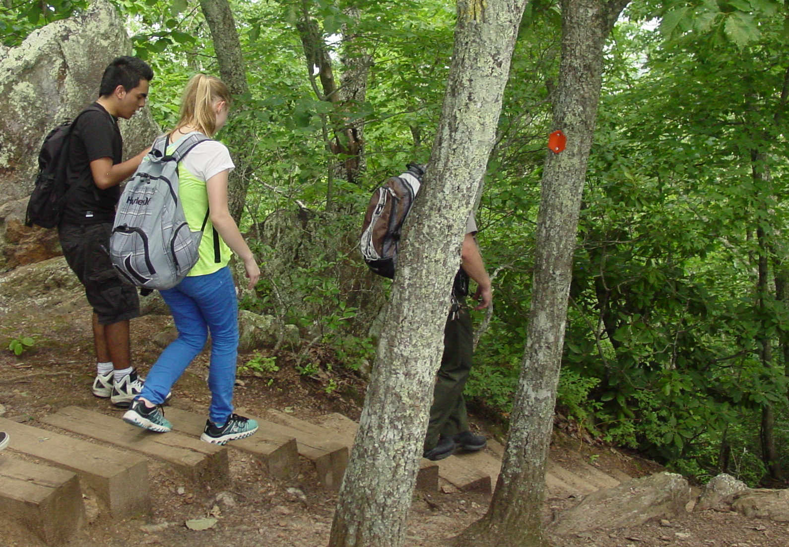
The Back Trail part of the loop includes some very steep steps - we were glad we did not come UP this way.
Many steps down And more steps..... Pause for a geology demonstration Mica-containing mineral used in makeup
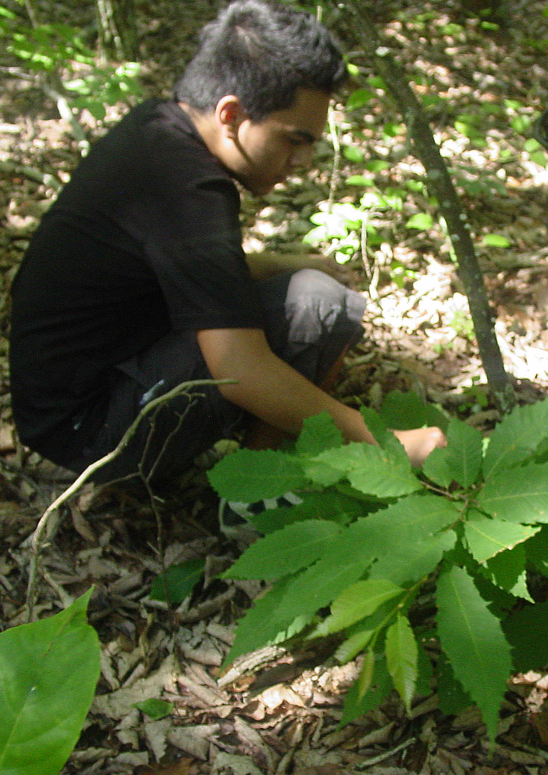
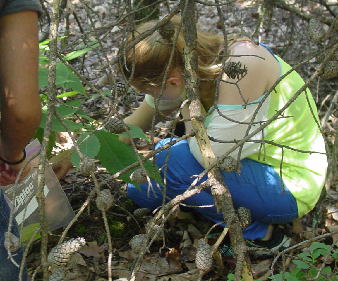
Going slightly off the main trail, samples were collected from shoots put out by blight-killed trees.
Willie collecting leaves Amara and partner bagging leaves Nicole and Maria verify sample labels Getting GPS coordinates
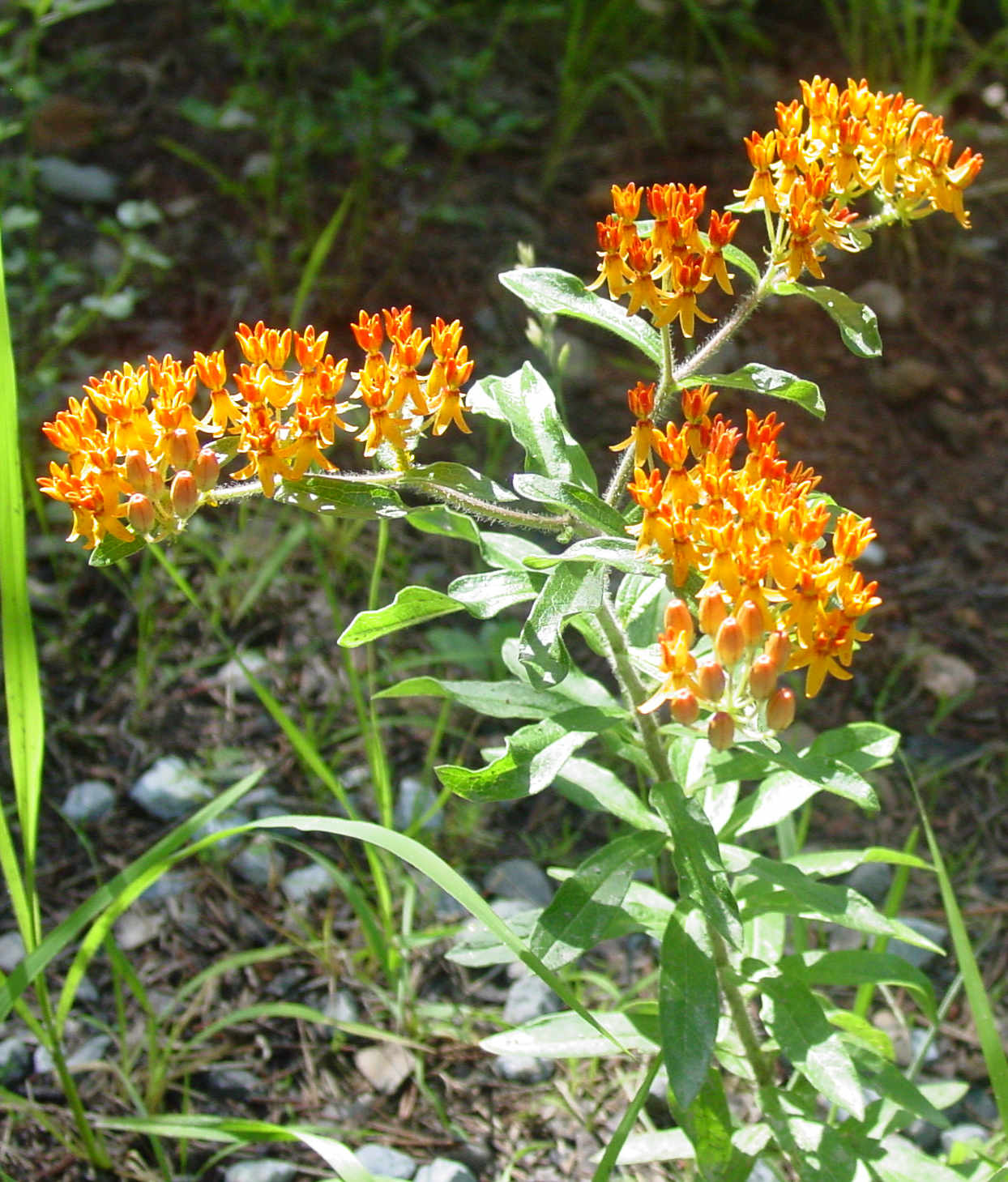
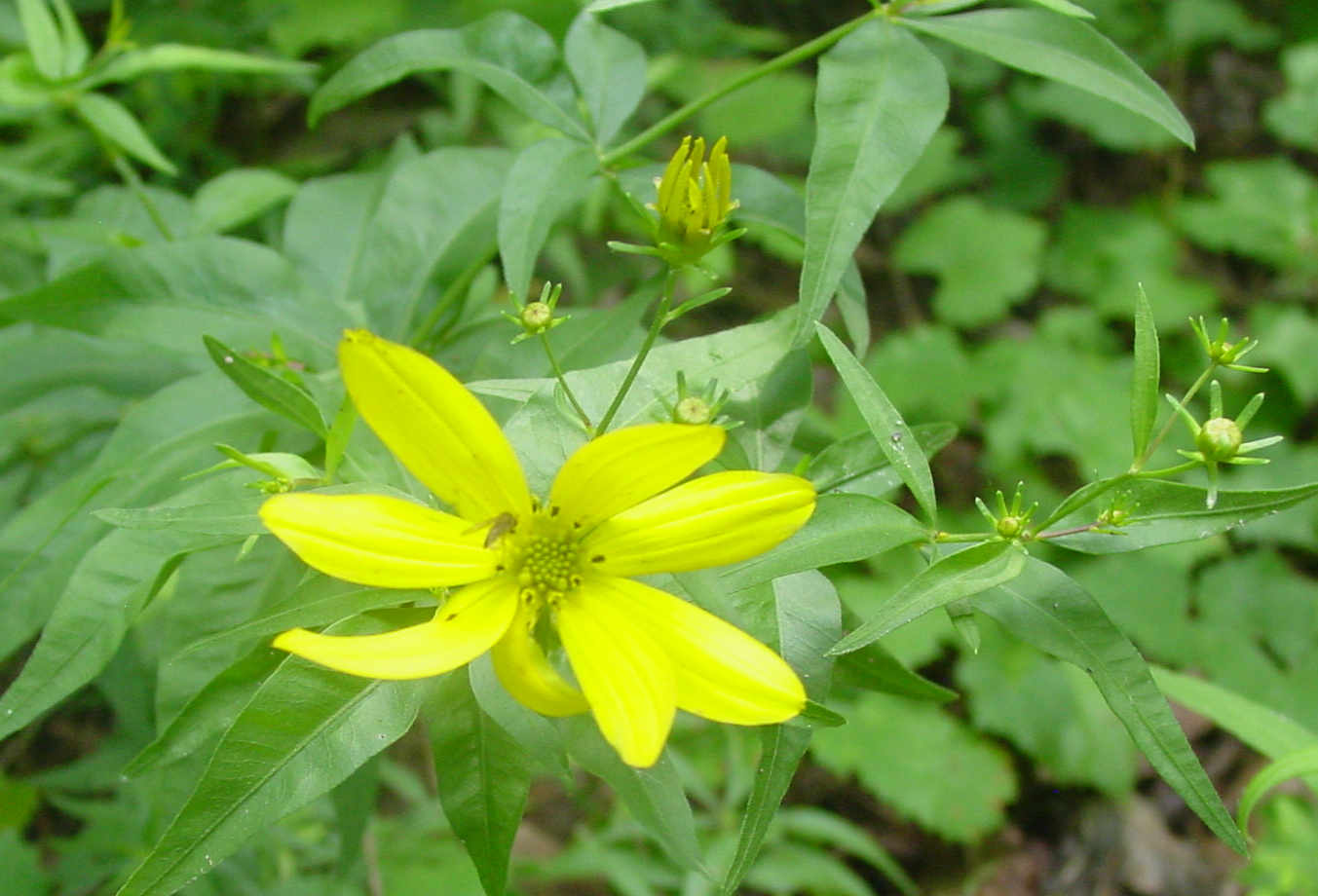
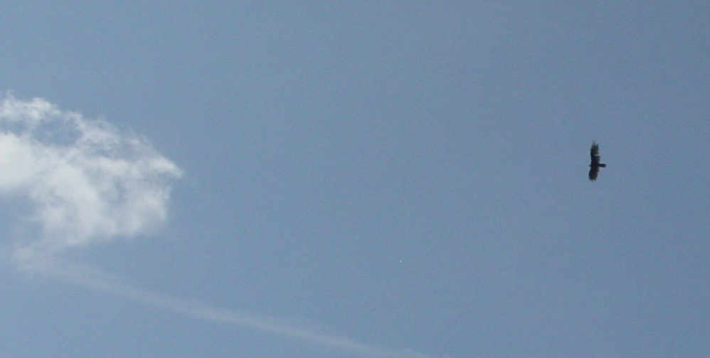
Our journalist was careful to capture some of the Biodiversity of the environment in which our samples exist.
Botanical forms Animal forms Spot the spider if you can
The final bit of trail to the parking lot.... and lunch.... and air conditioning!
The pace picked up here. Friday, June 13 th, 2014. The B3 Summer Science Camp Blog
Our second and equally important field trip took place today. Ladies and gentlemen, may I present to you the star students and staff
who took the challenge of hiking about a million miles around Crowder's Mountain (yup, you heard it right)... the B-3 champs! Under the leadership
of Ranger Kelly Cook, we were able to locate several of the native American chestnut trees and collect samples extremely vital for our work in the lab next week.
We were not only able to collect samples, but we also learned about the ways nature depends on occasional wildfires. Between watching Ranger Kelly
consume wild berries sporadically and having the opportunity to capture so many beautiful images (with both Dr.Weller's camera and my eyes), I am most
definitely glad we were able to take an enormous hike and explore the wonders earth contains.
----- Annabel Richards
June 16 th
The Lab Work Begins
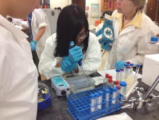
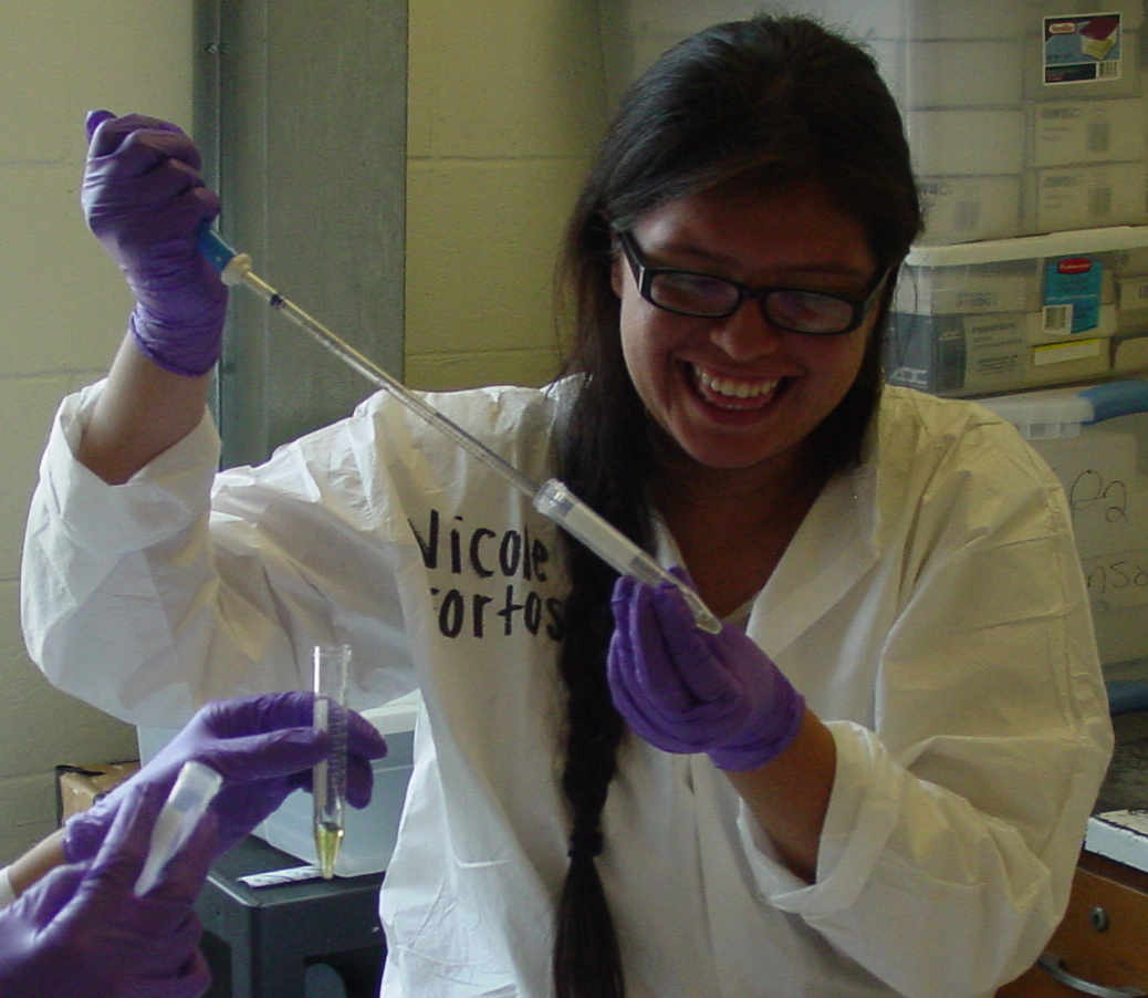
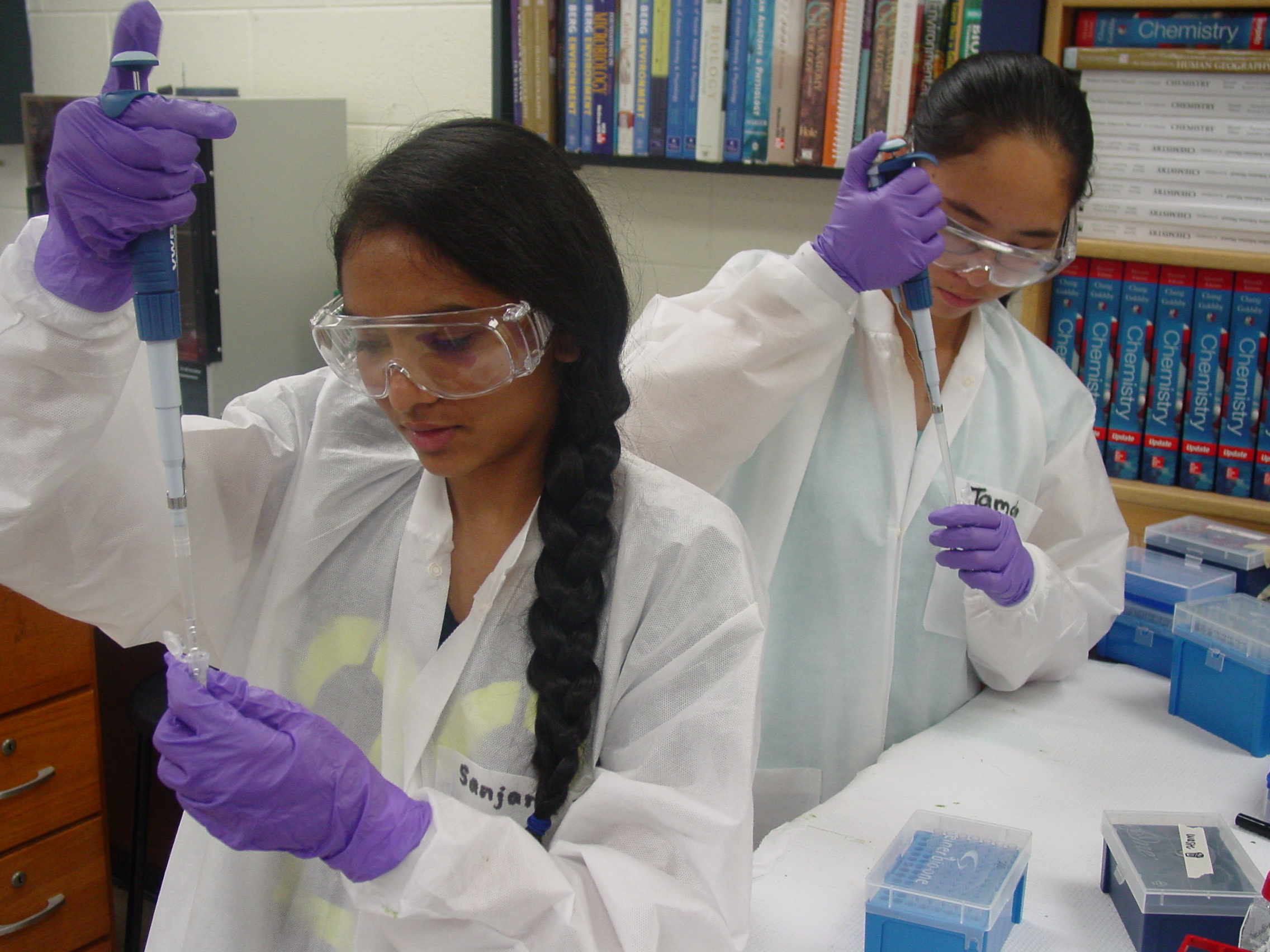
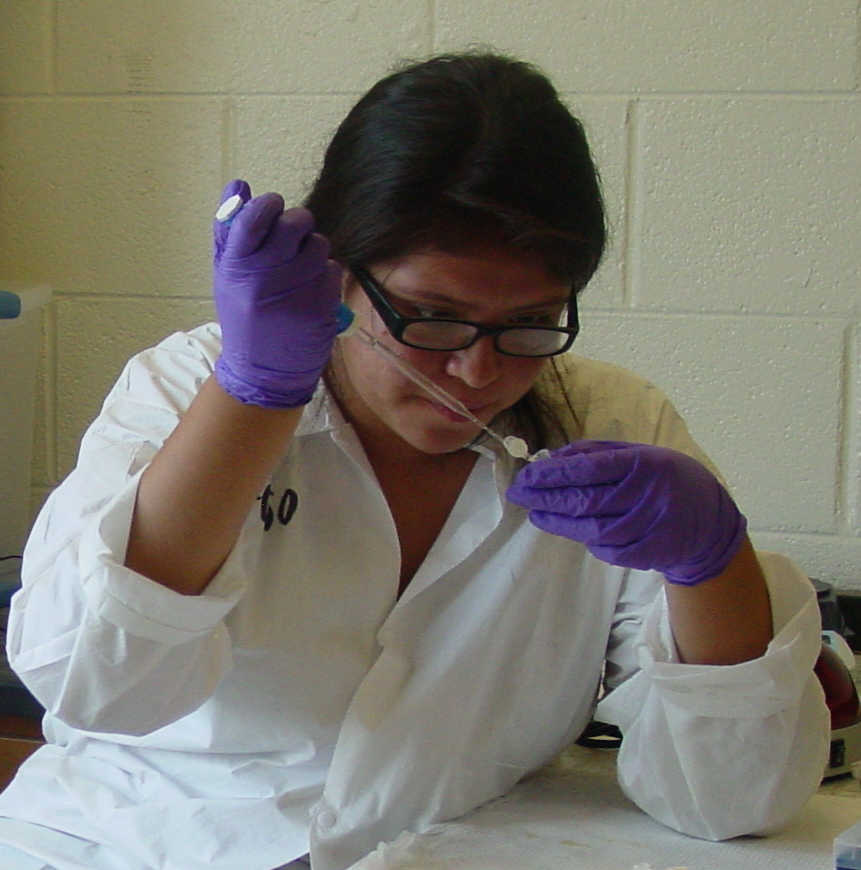
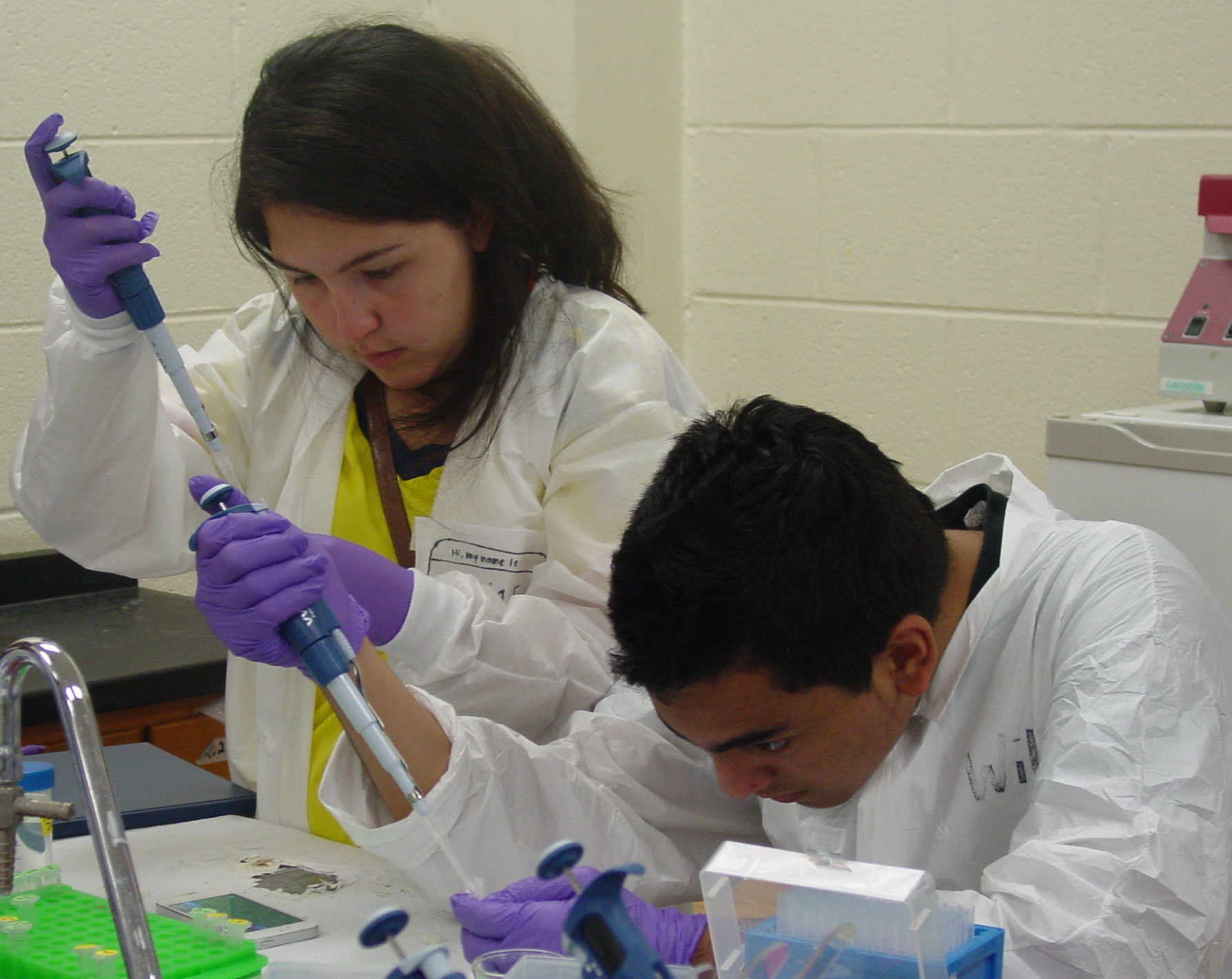
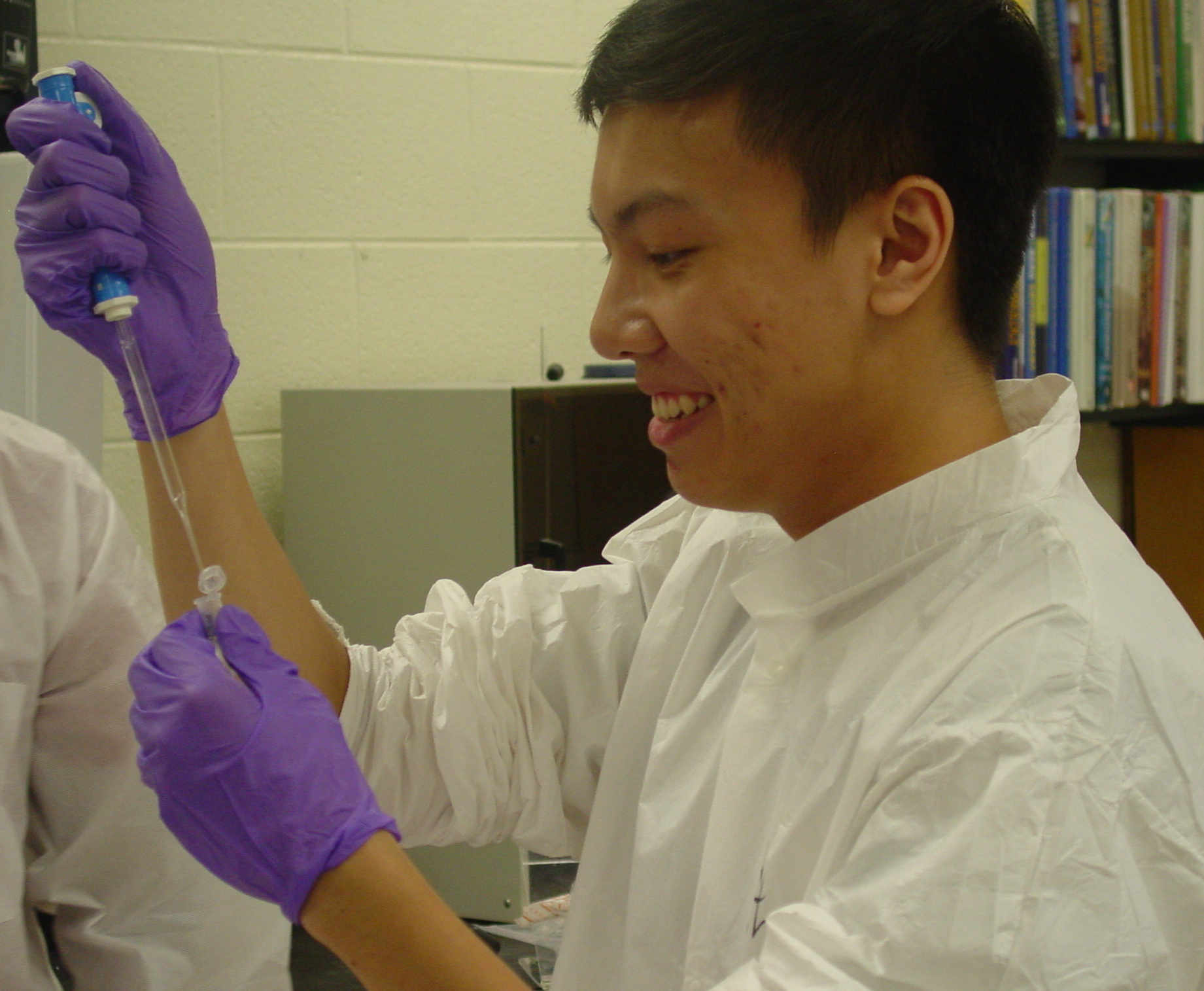
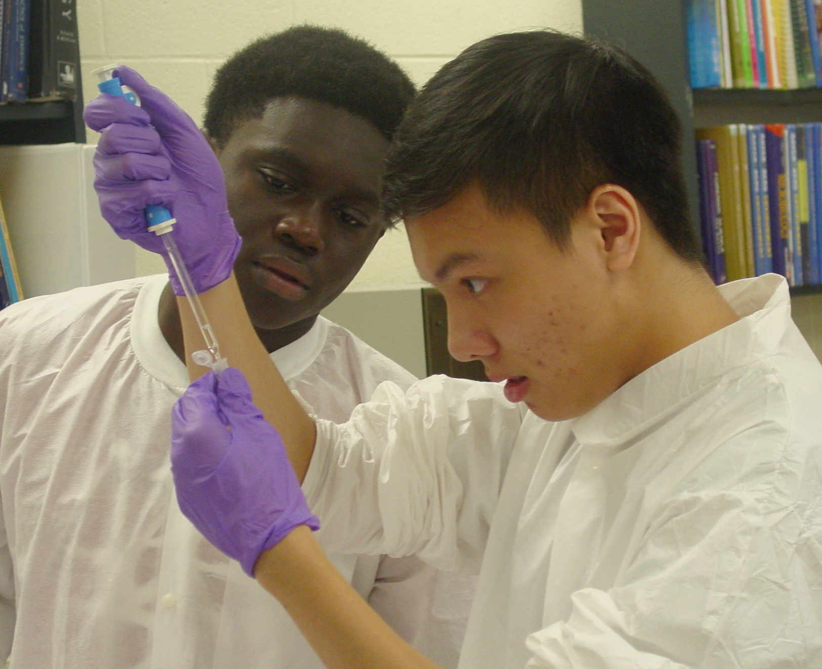
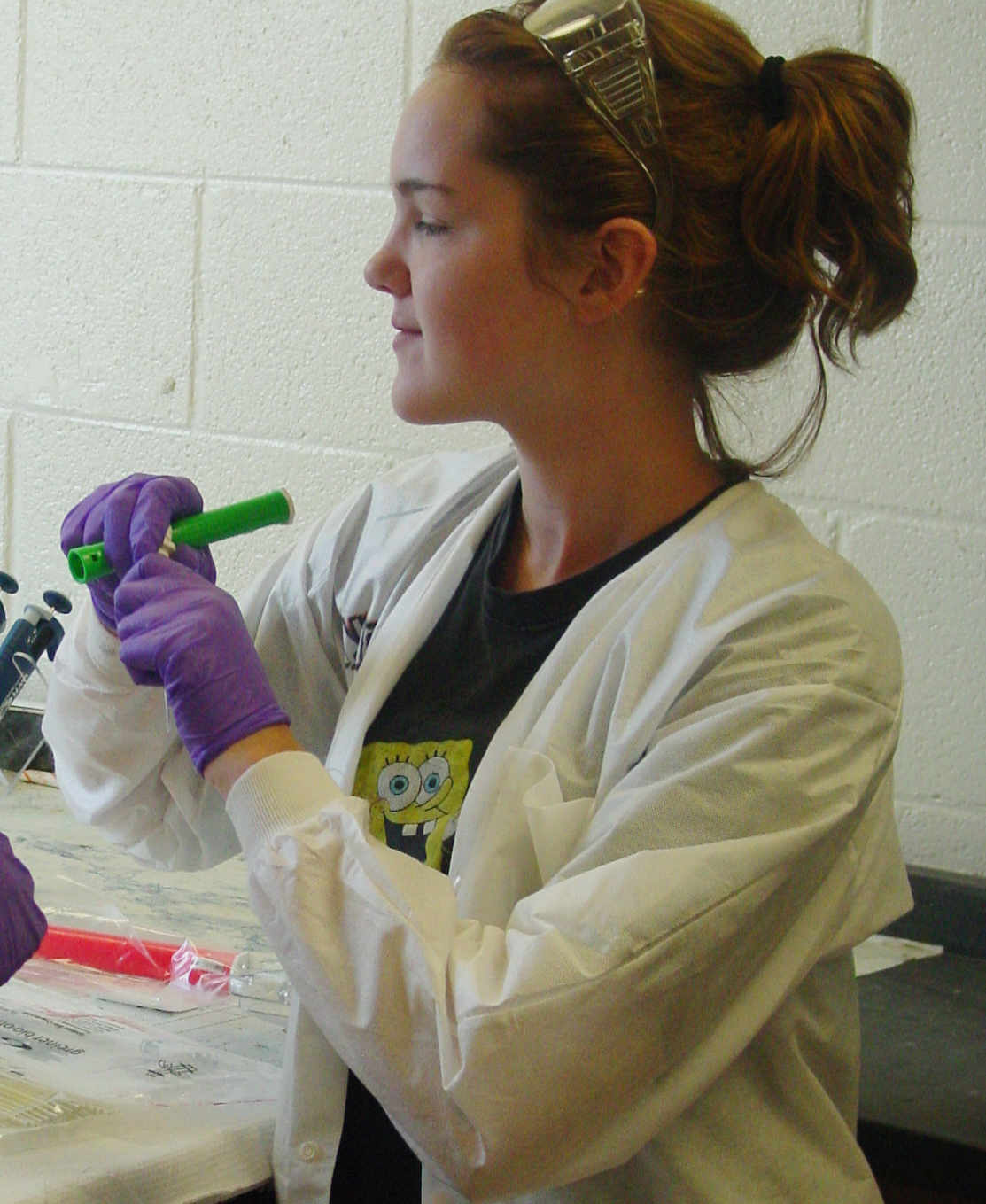
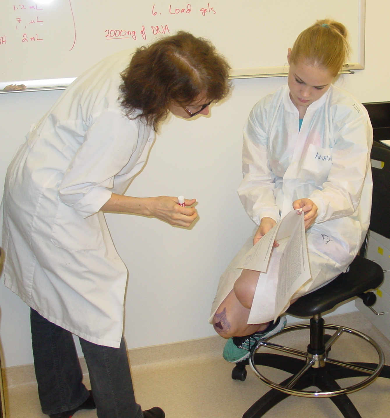
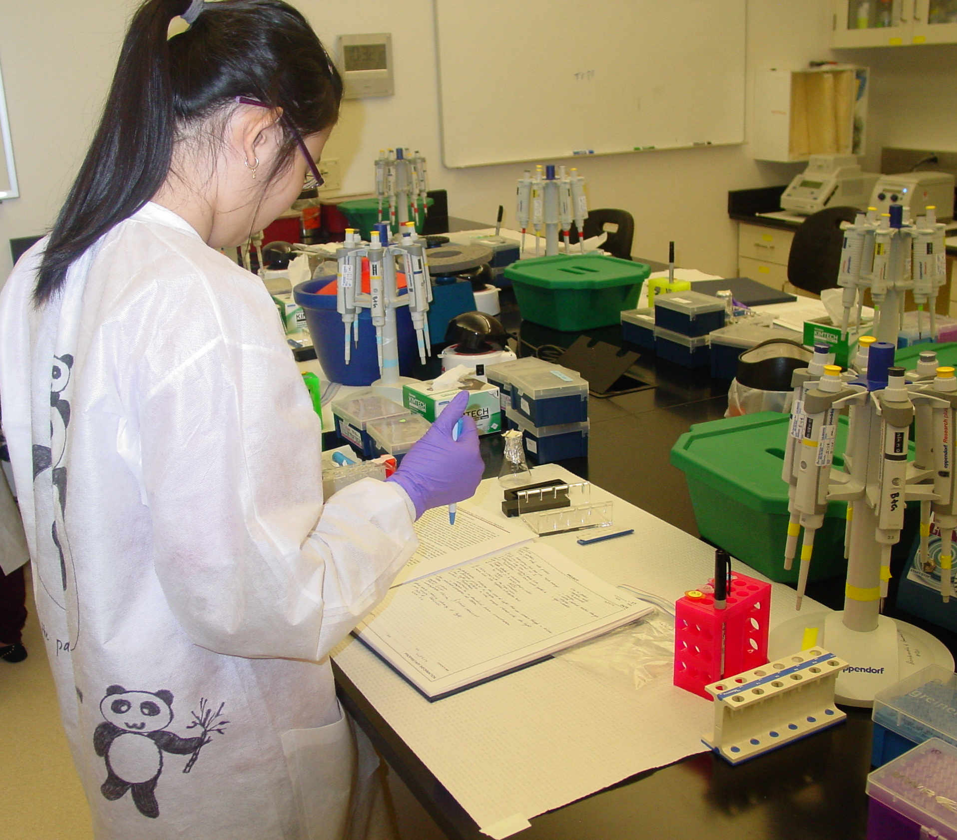
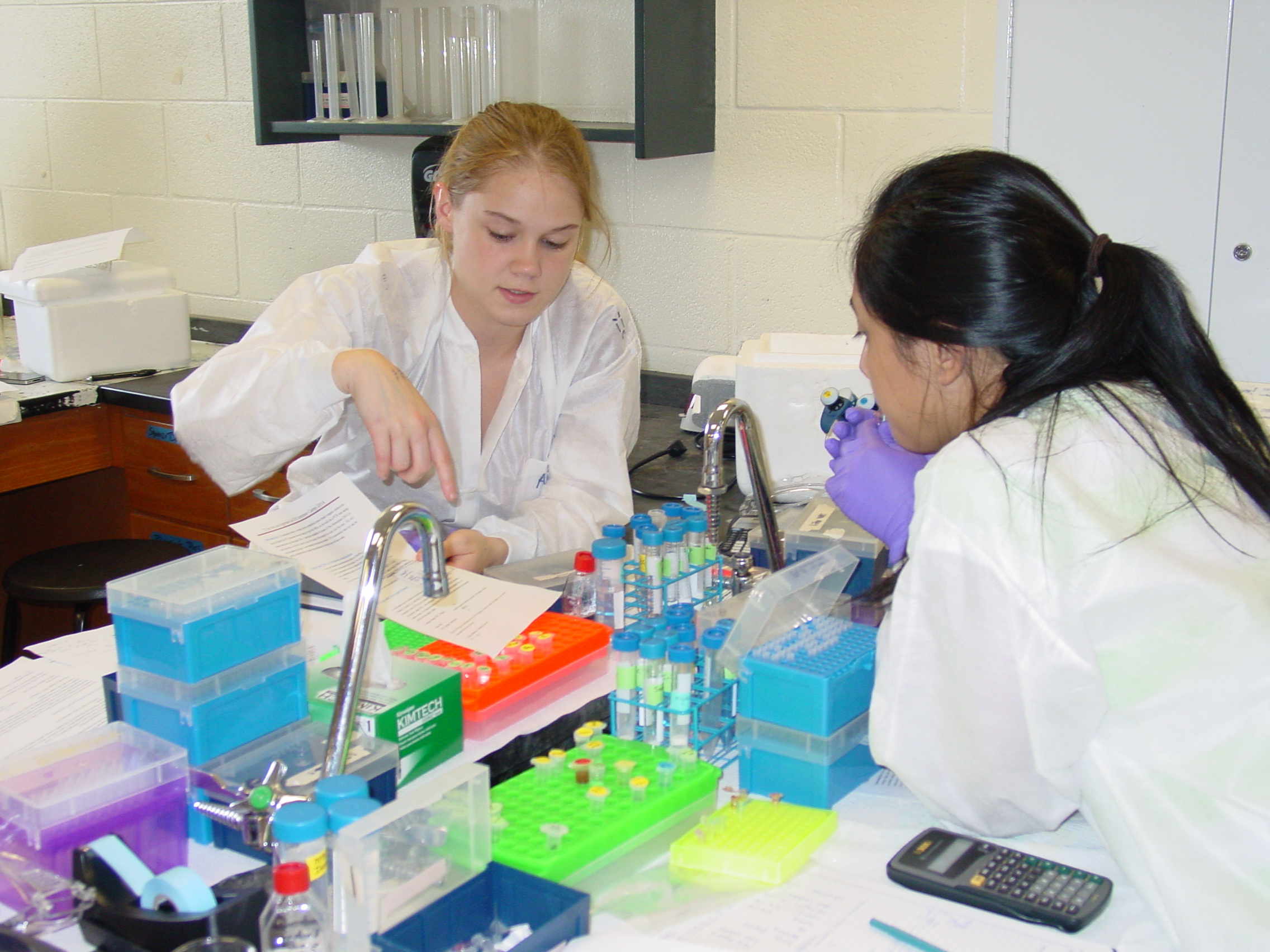
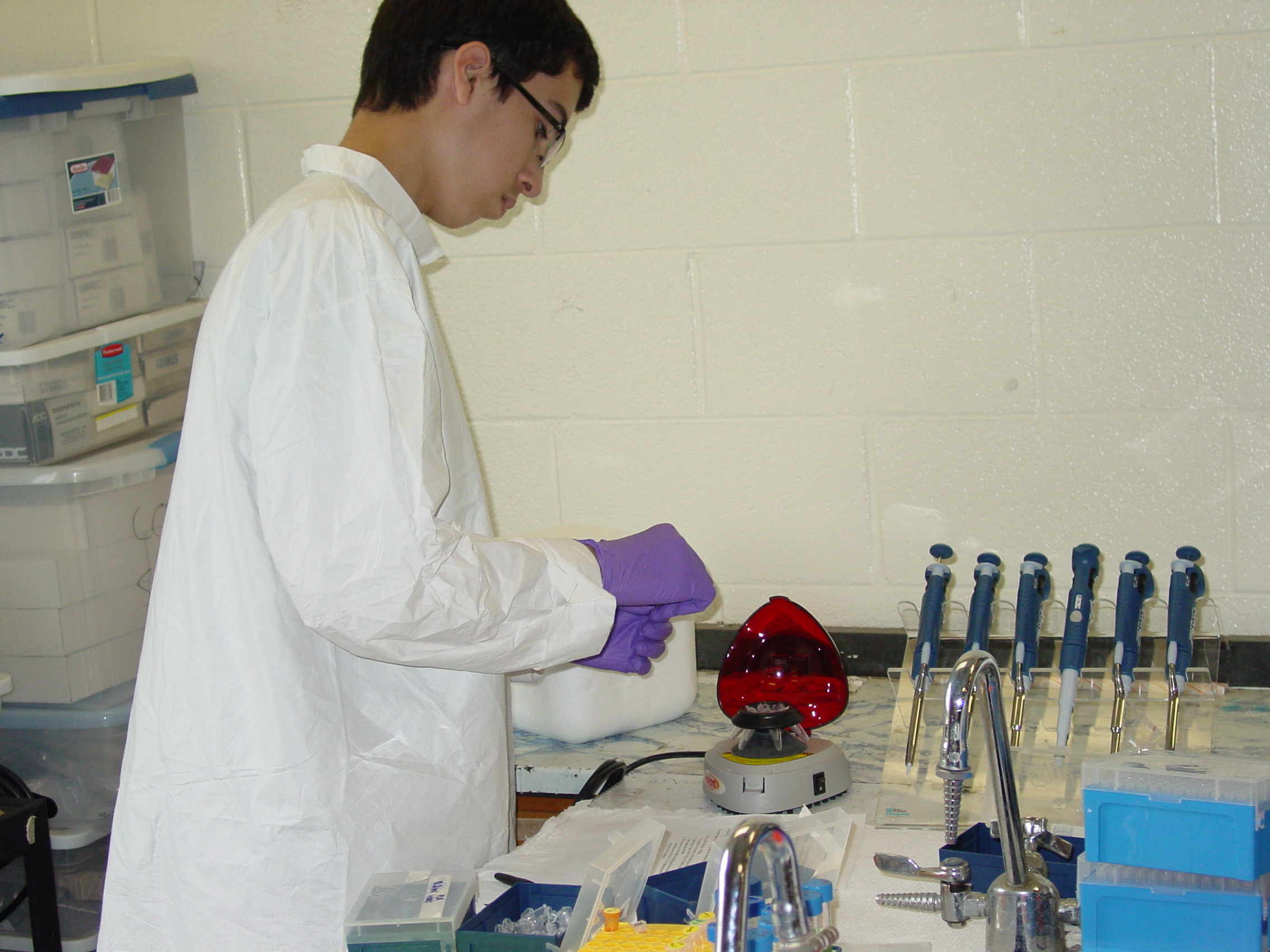
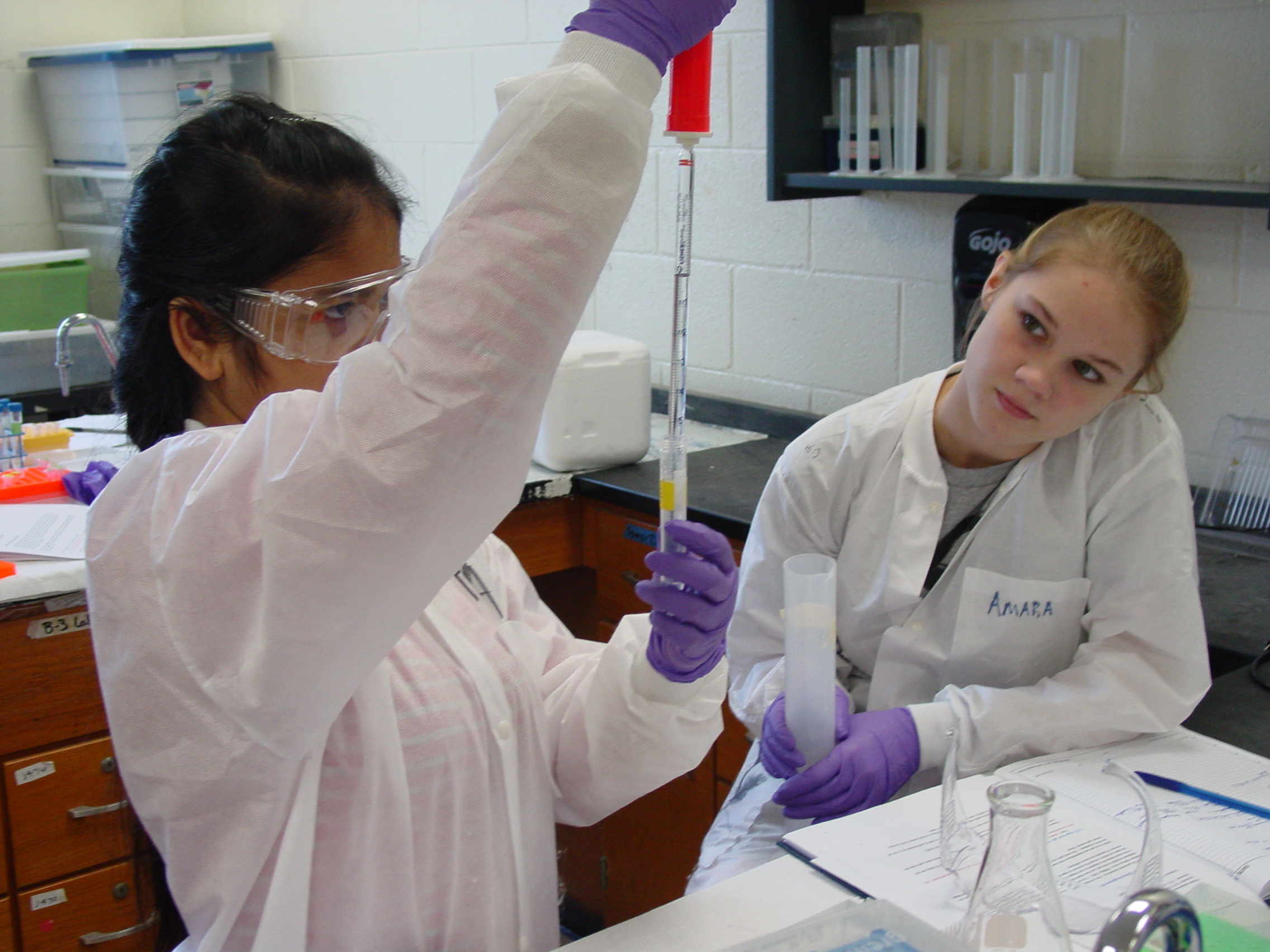
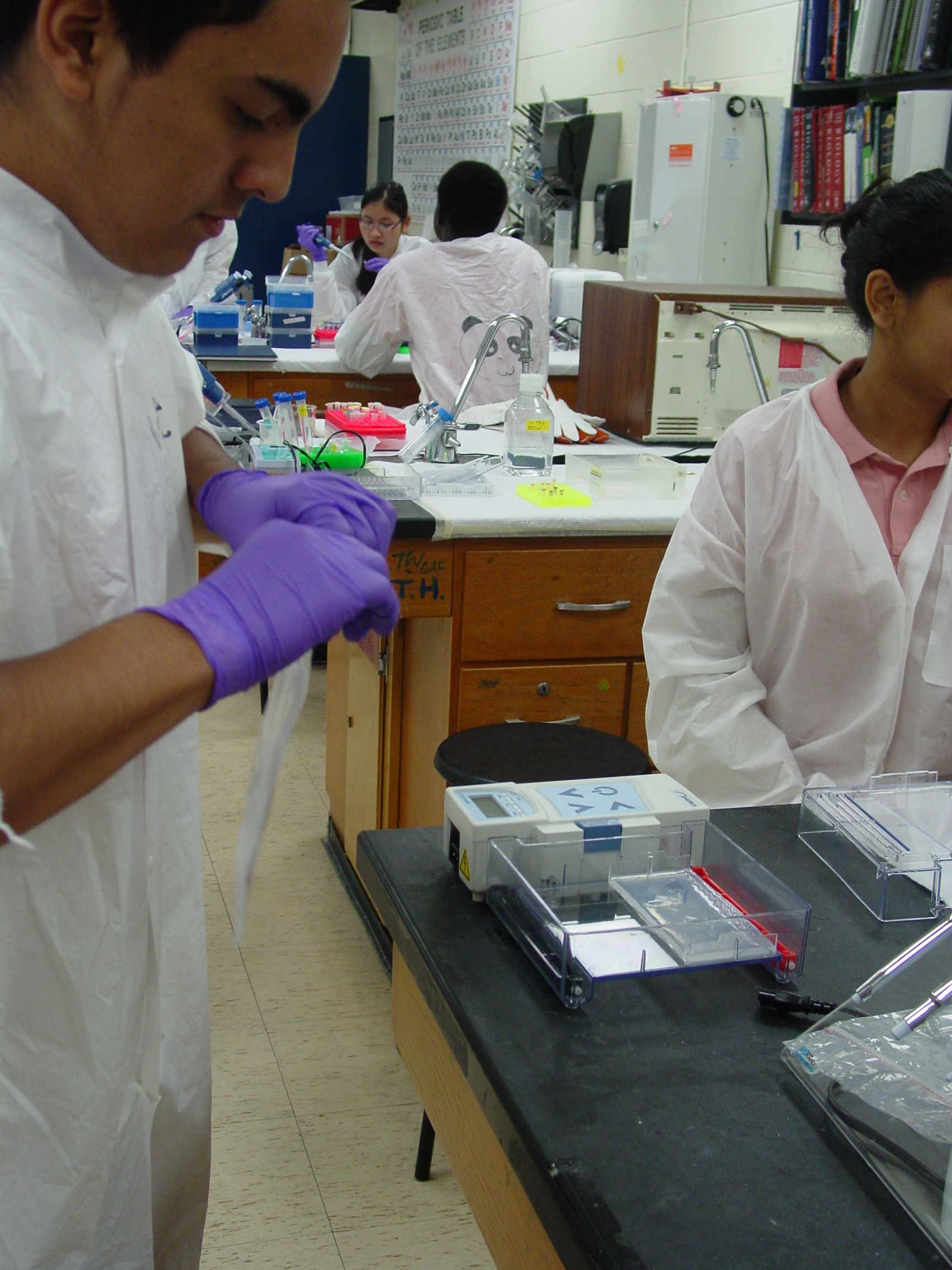
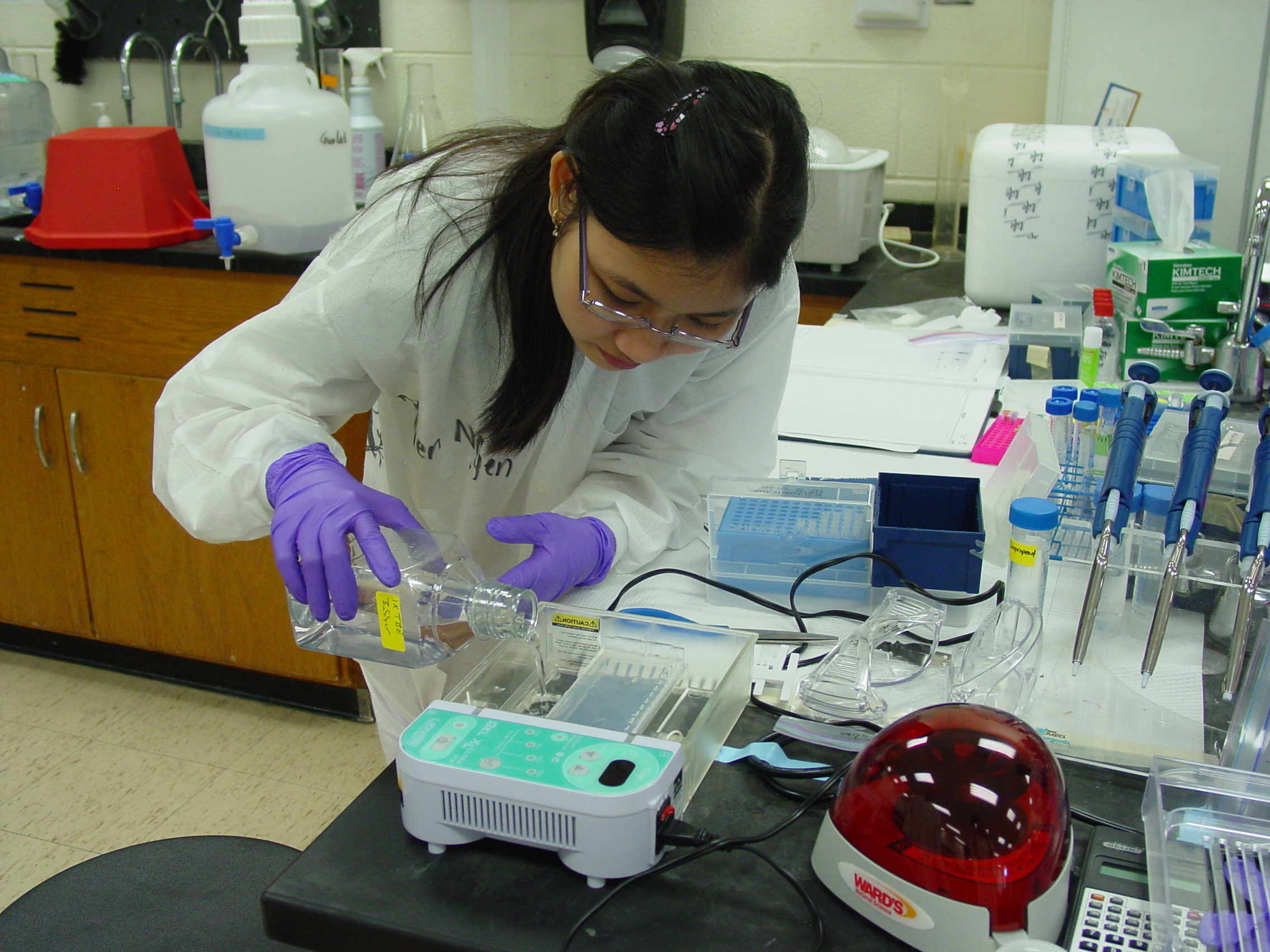
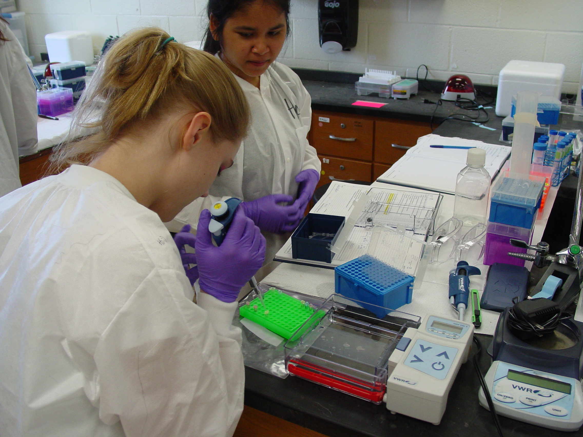
We used today to refresh (or learn) basic molecular biology skills, including pipetting with micropipetters and serological pipettes and using microbalances.
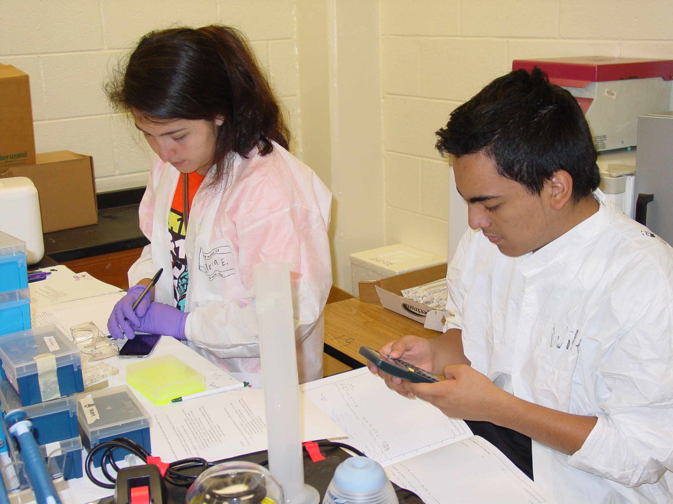
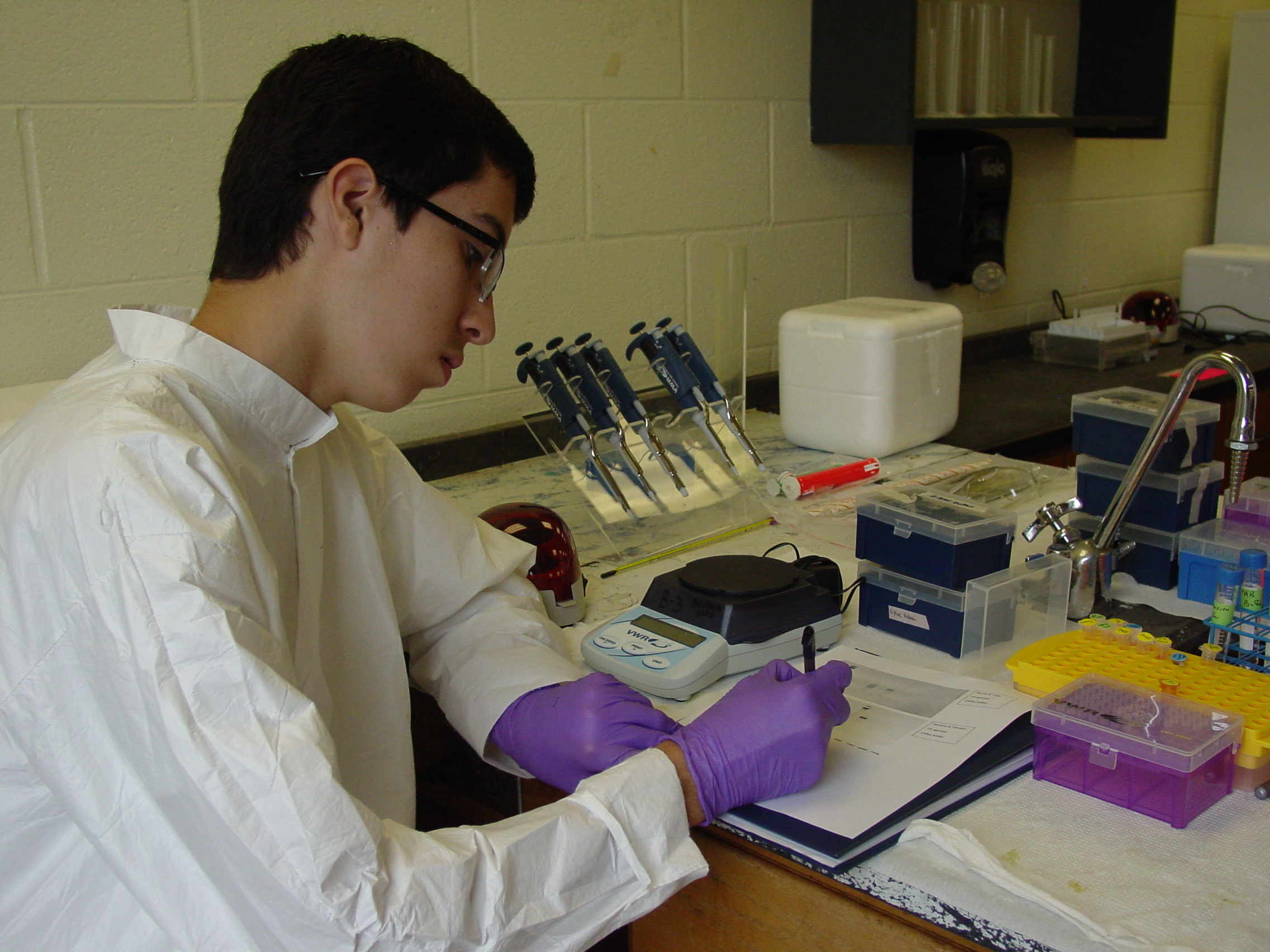
Recording measurements and observations and then analyzing the data is just as important.
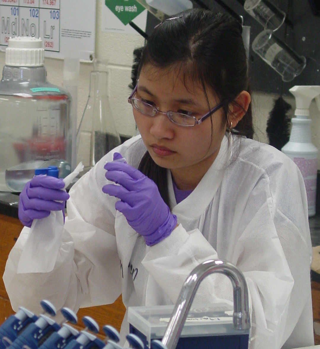
Each team worked out its own approach, some trading off measuring while the partner recorded and others preferring to record their own measurements.
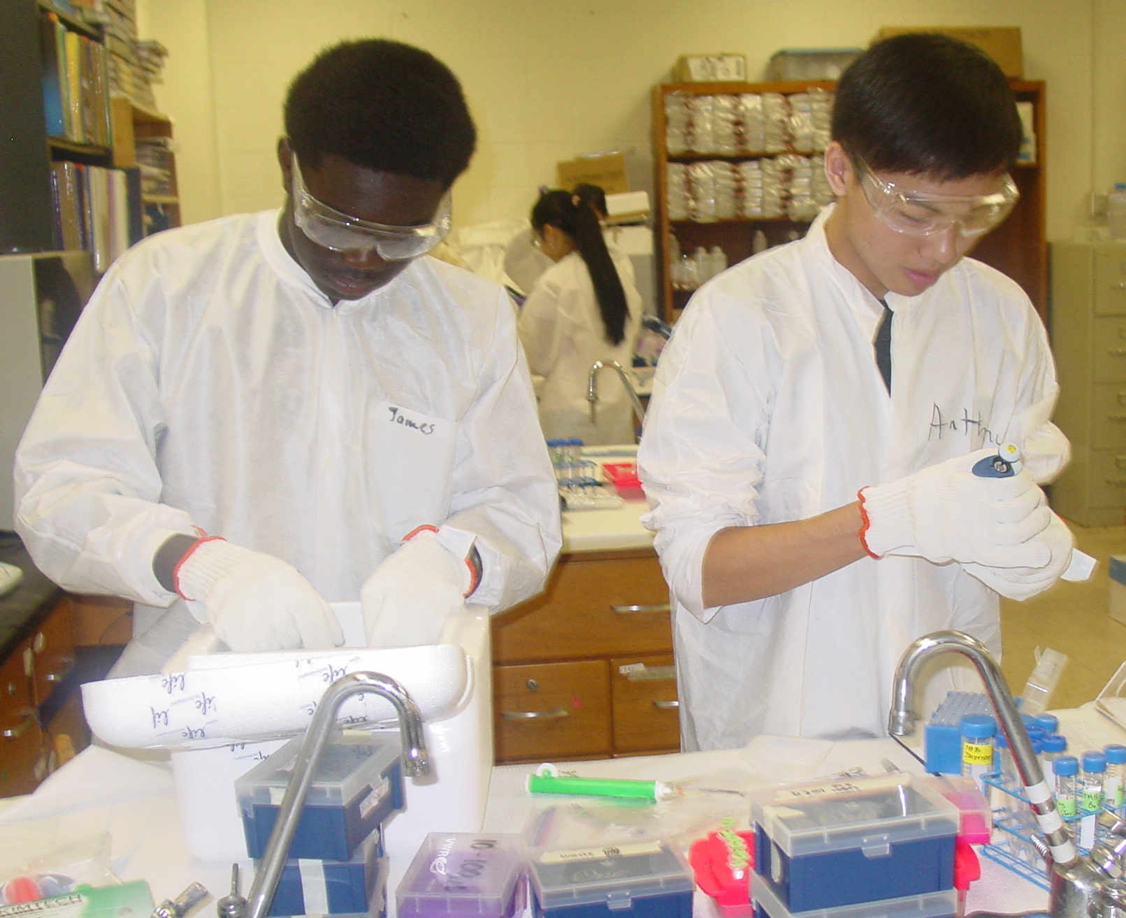
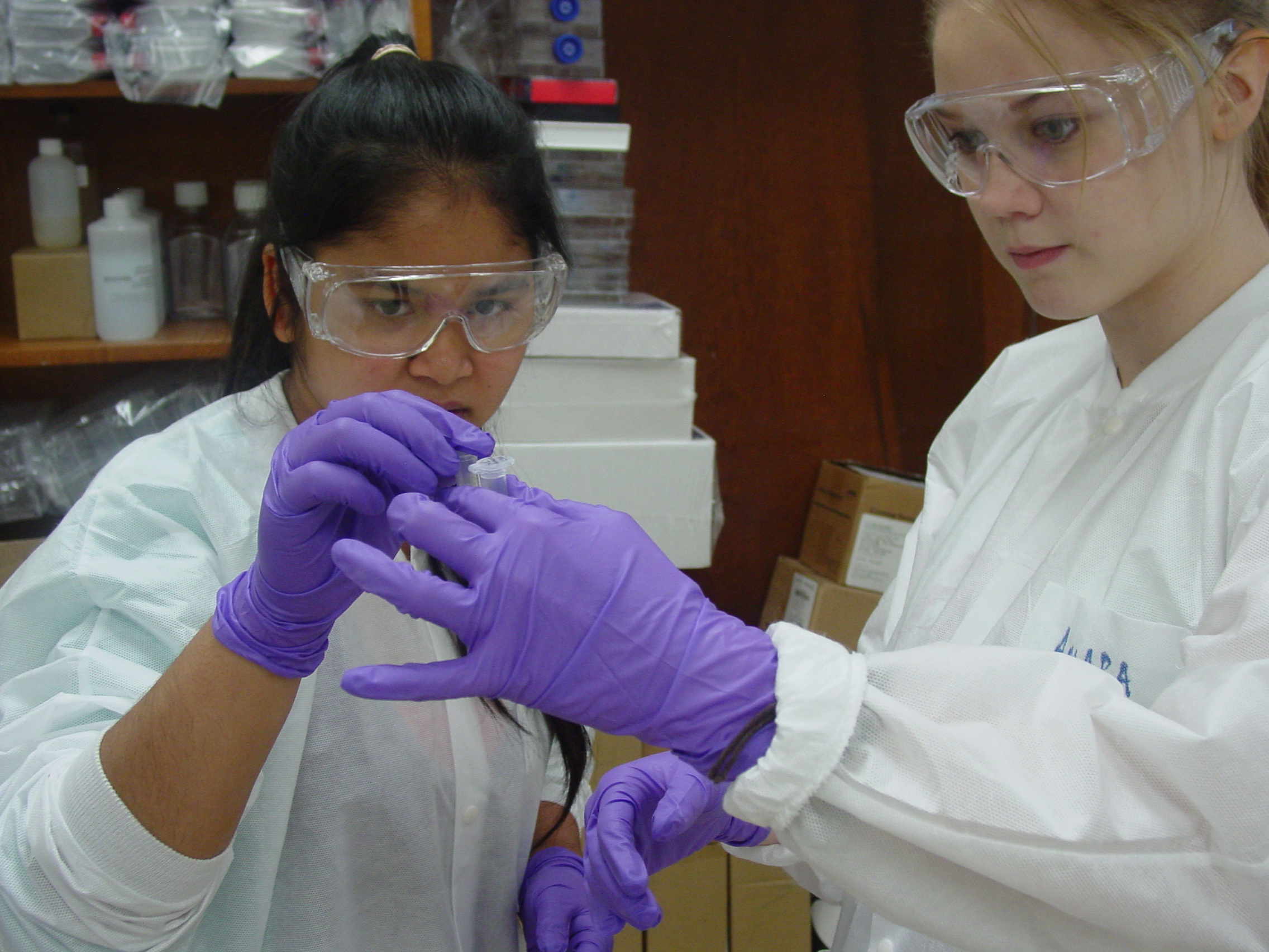
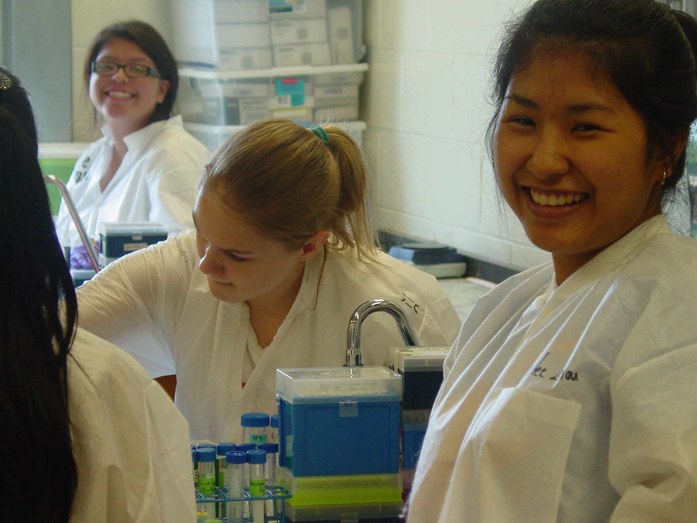
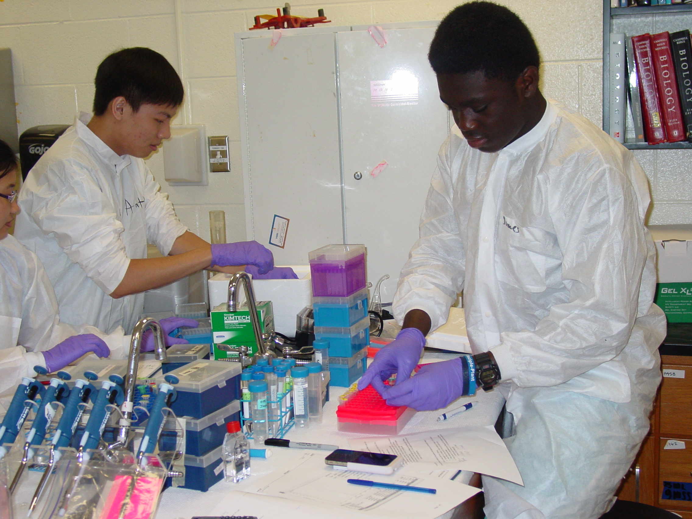
These pictures give a feeling for fully kitted-out benches, and students hard at work.
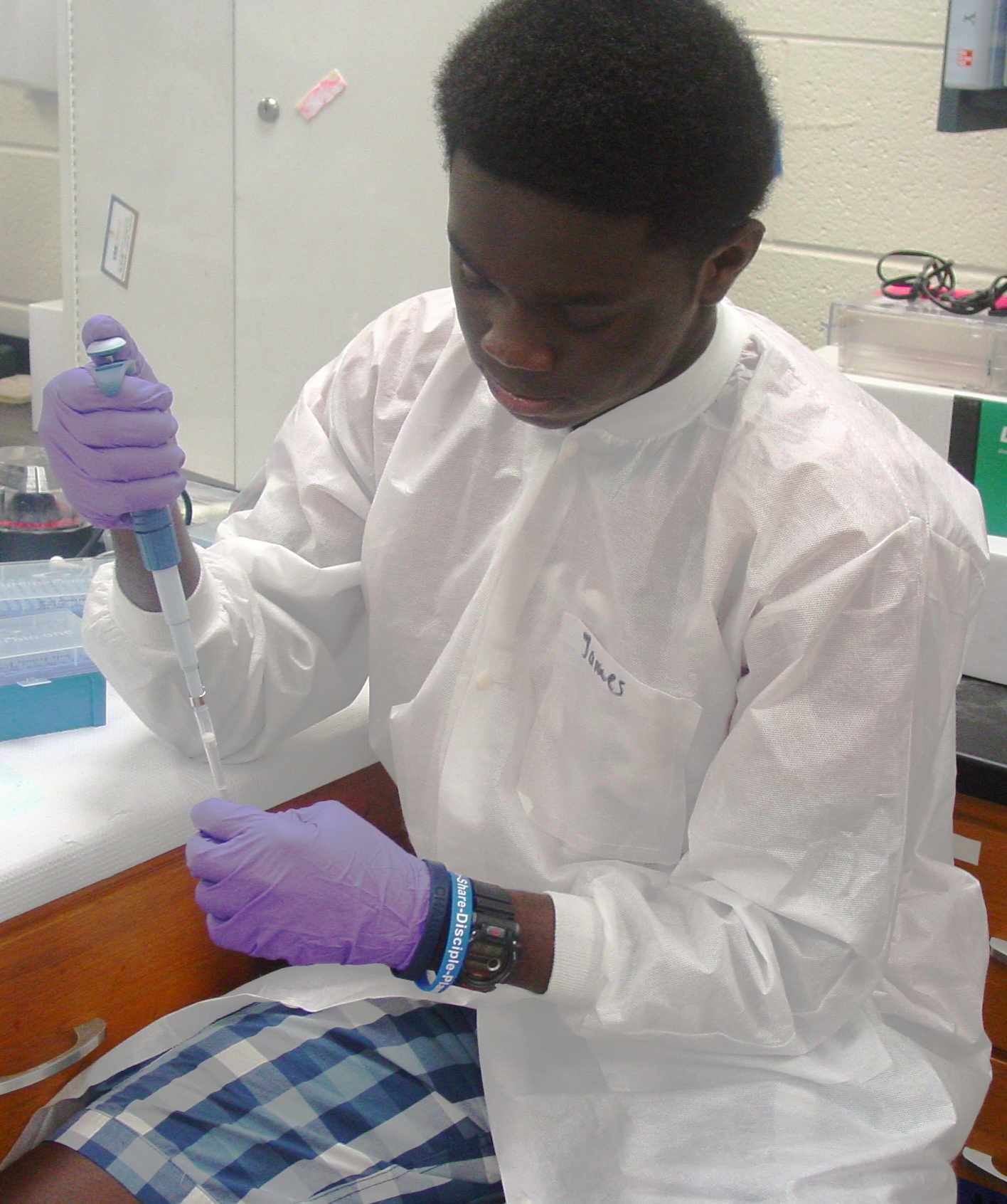
Benches with Materials in Place. Ms.Smith Reviews the Protocol with Nhi and Amara Anthony on Measurement Detail Duyen Recording the data. James on Measurement Detail Annabel, Nicole, Doudgie and Tripp consider their options Think It'll Work? Willie and Maria focusing Sanjana - is this precise or accurate? Amara and Hoa starting up. Monday, June 16 th, 2014. The B3 Summer Science Camp Blog
This week in the lab is planned to include a ton of exciting work. Today, students practiced skills important for a successful lab environment
to prepare for a smooth experience throughout the week. We learned the names of a few lab equipment items that we are soon to be familiar with, and we
practiced the proper methods to use them. Practice using these items began with water, and then we moved on to something denser: glycerol. Our two main
tools were micropipettes and serological pipettes. For every measurement, we had to follow specific steps for weighing the portions and calculating
out actual results in comparison to the results were trying to receive. It was within this practice, that students were taught the difference between
the words "precise" and "accurate." Today would be classified as the basic introduction to the LAB, or, everything we are soon to
be familiar with throughout the next few days.
----- Annabel Richards
June 17 th
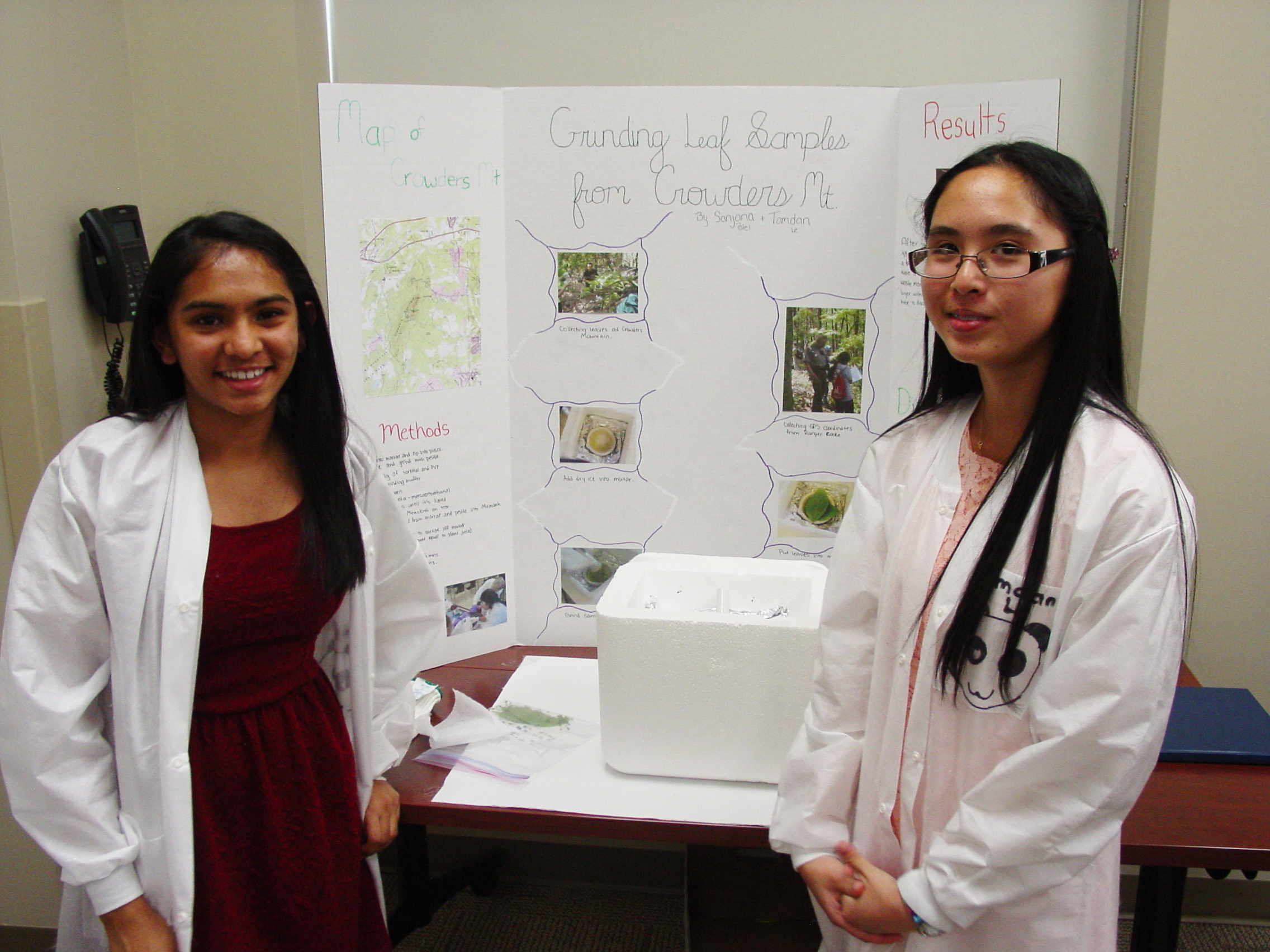
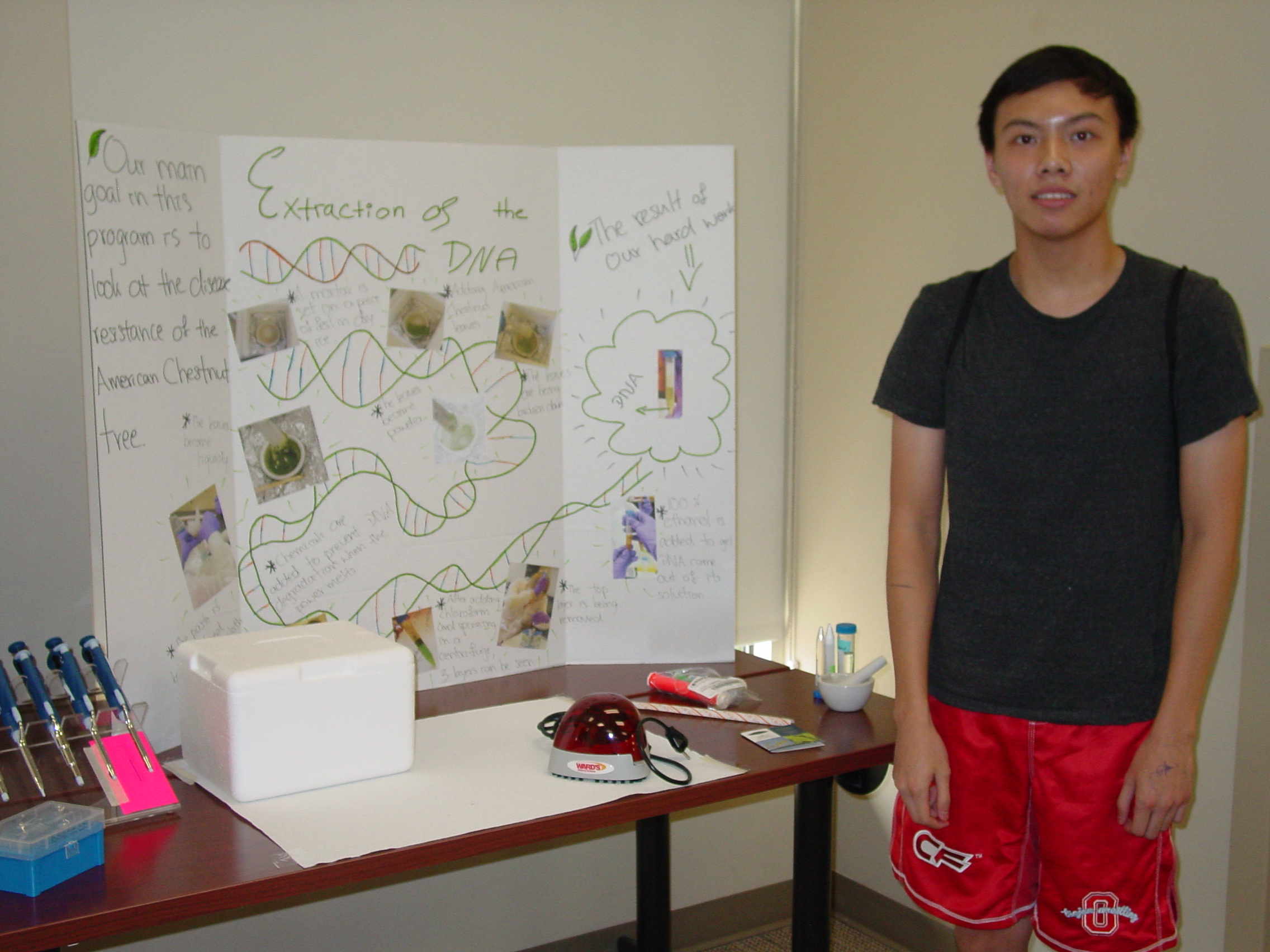
DNA Purification from Leaf Material
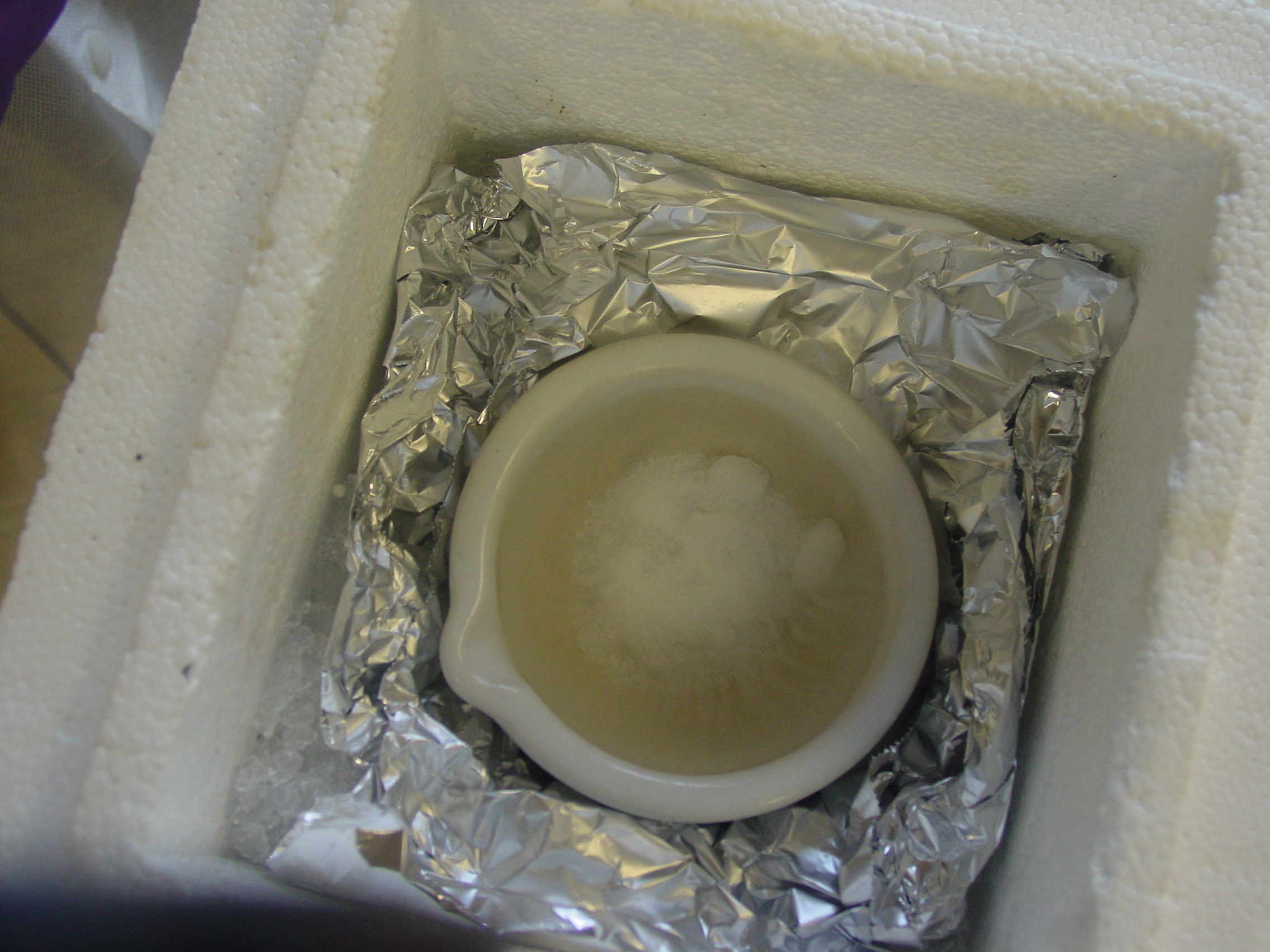
In order to get the DNA from cells, we have to break them open.
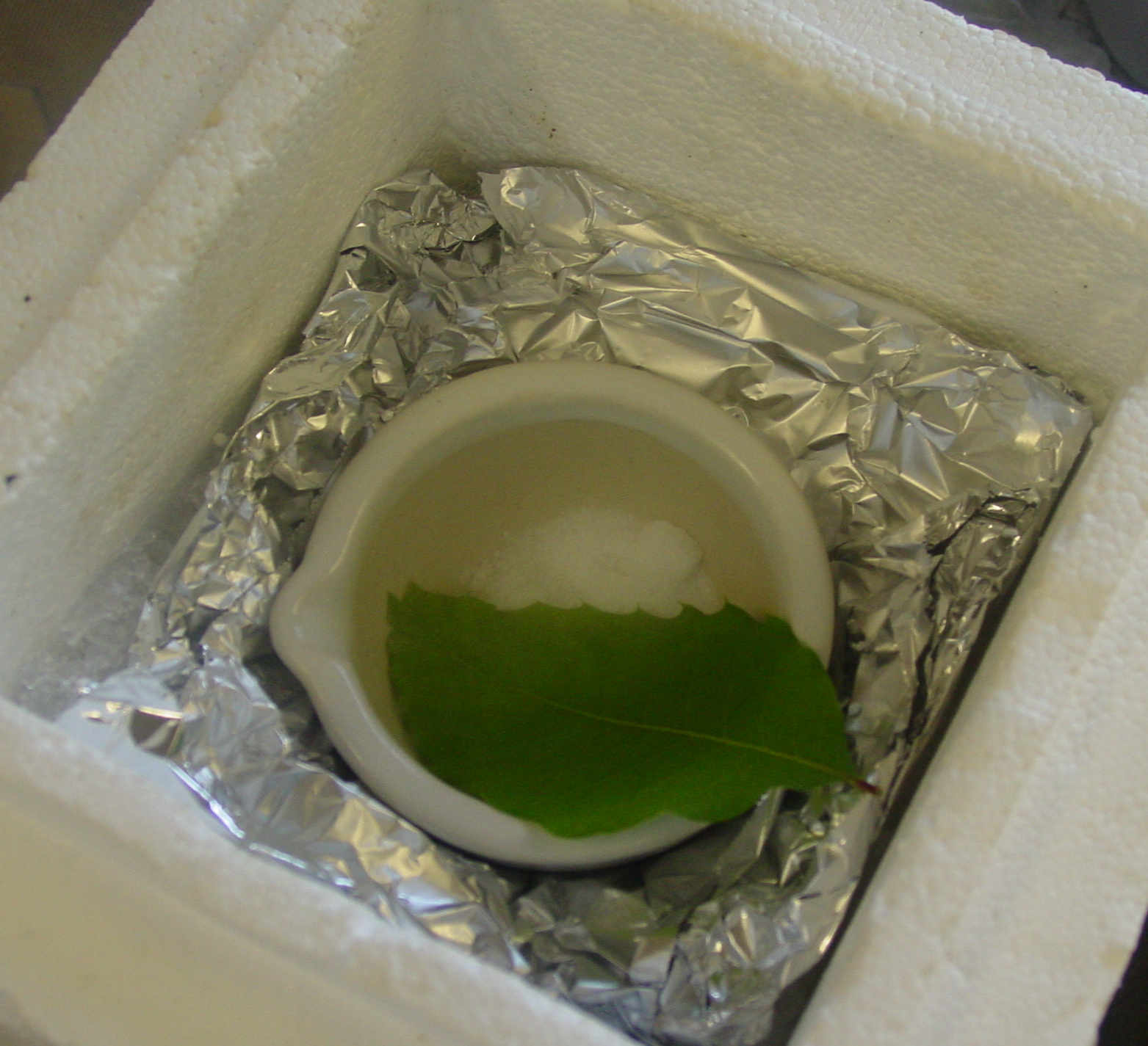
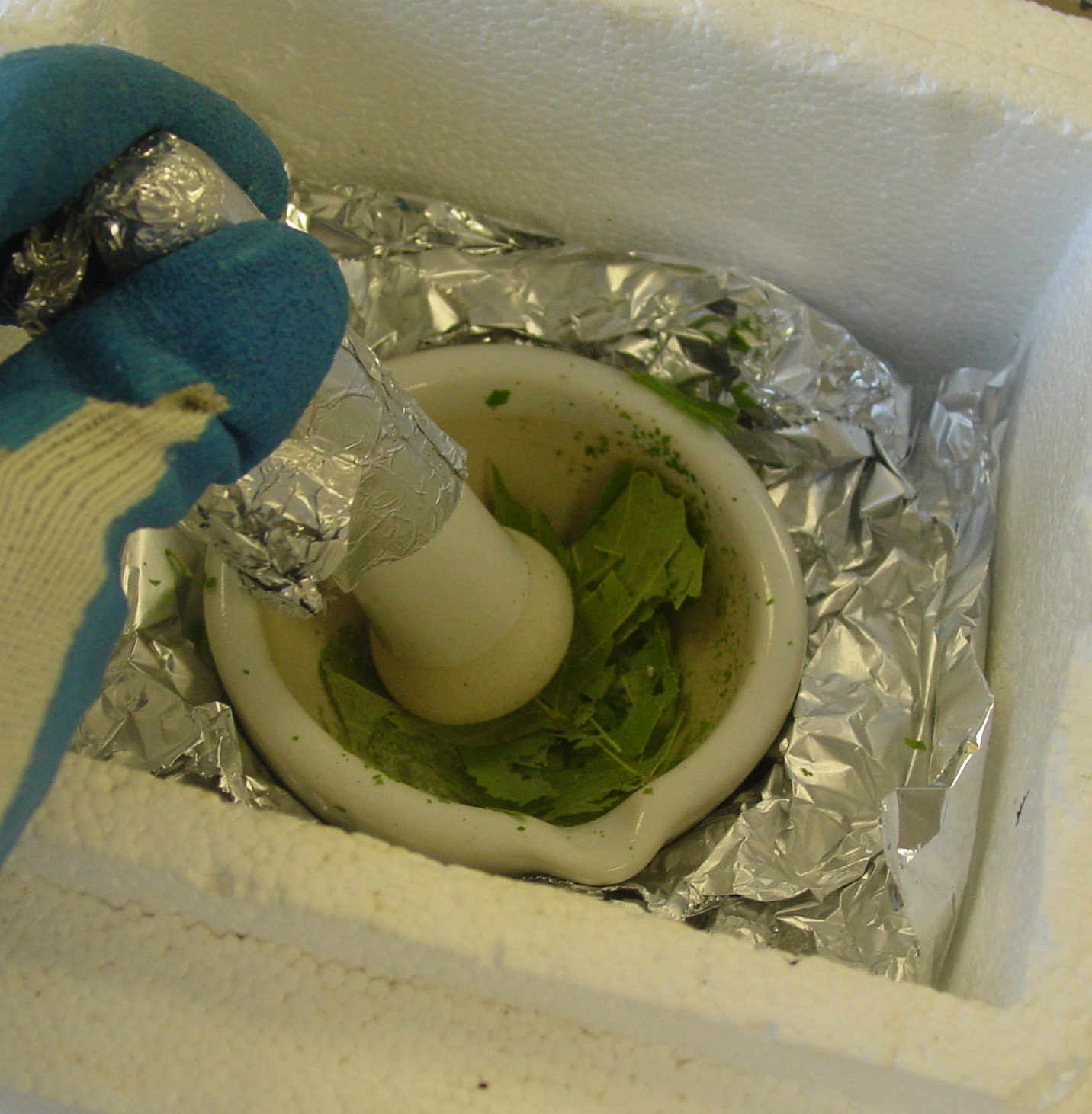
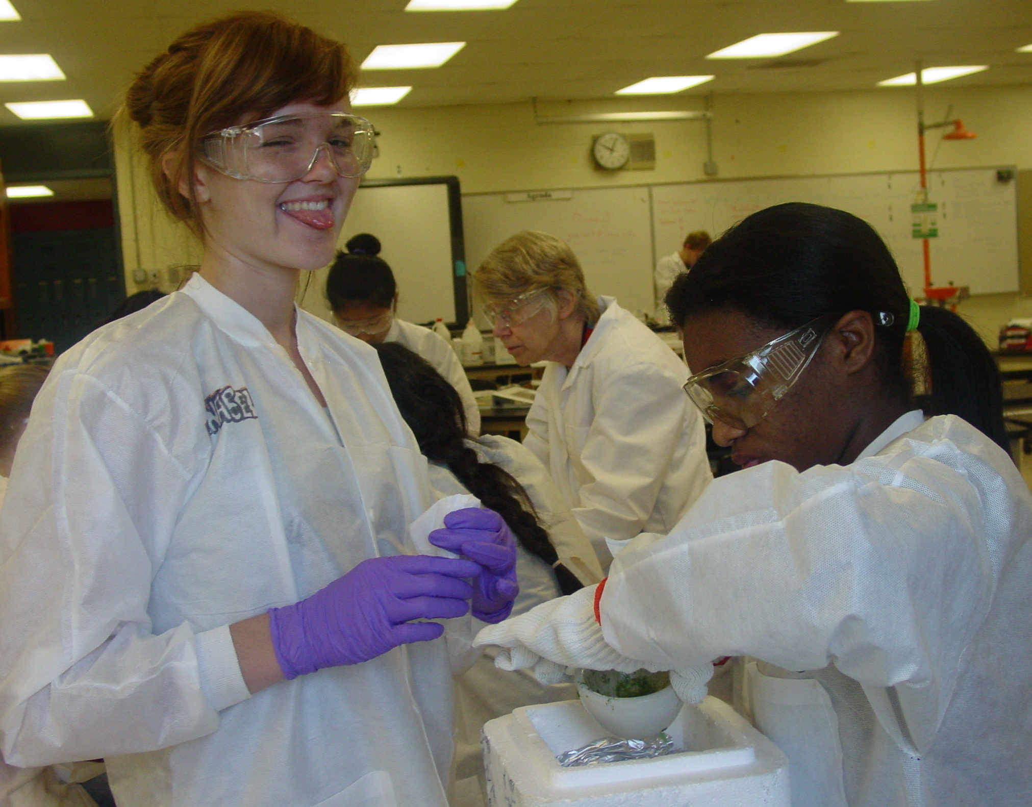
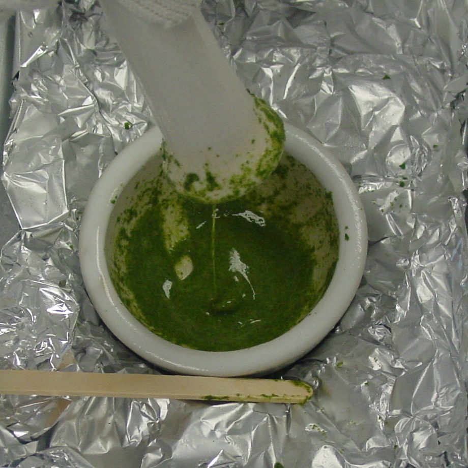
Plant cells have really tough cell walls, so we grind them using dry ice in mortars and pestles.
We include lots of chemicals to keep the plant cell contents from modifying or breaking down the DNA.
A mortar is set on a piece of foil in wet ice, and dry ice is put inside it, and then some leaves.
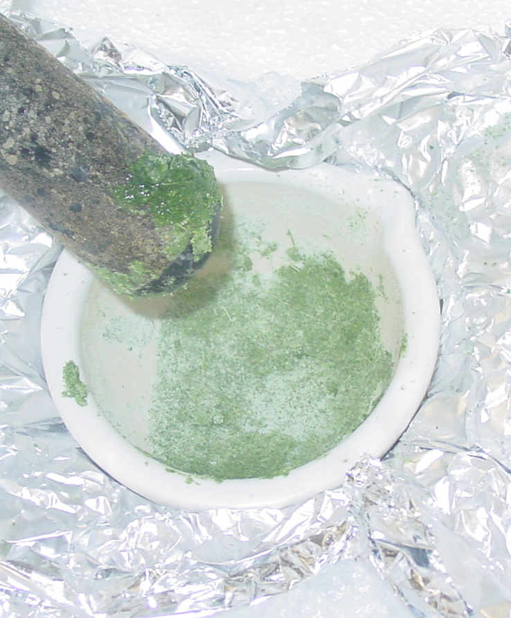
The leaves are broken up and then ground to a dry powder.
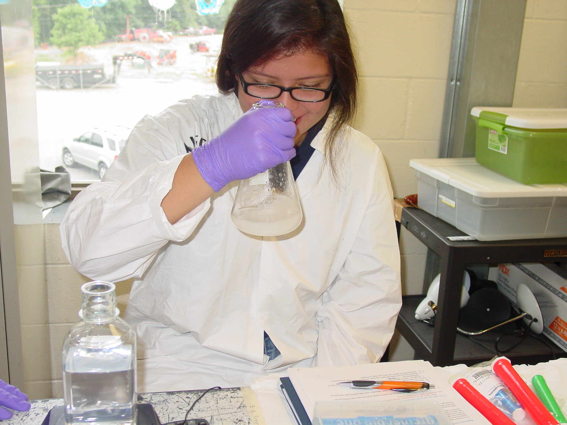
Chemicals are added to prevent DNA degradation when the powder melts: teamwork is required.
Some of the chemicals smell really bad (beta-mercaptoethanol).
The paste becomes quite liquid and is filtered through Miracloth into a tube.
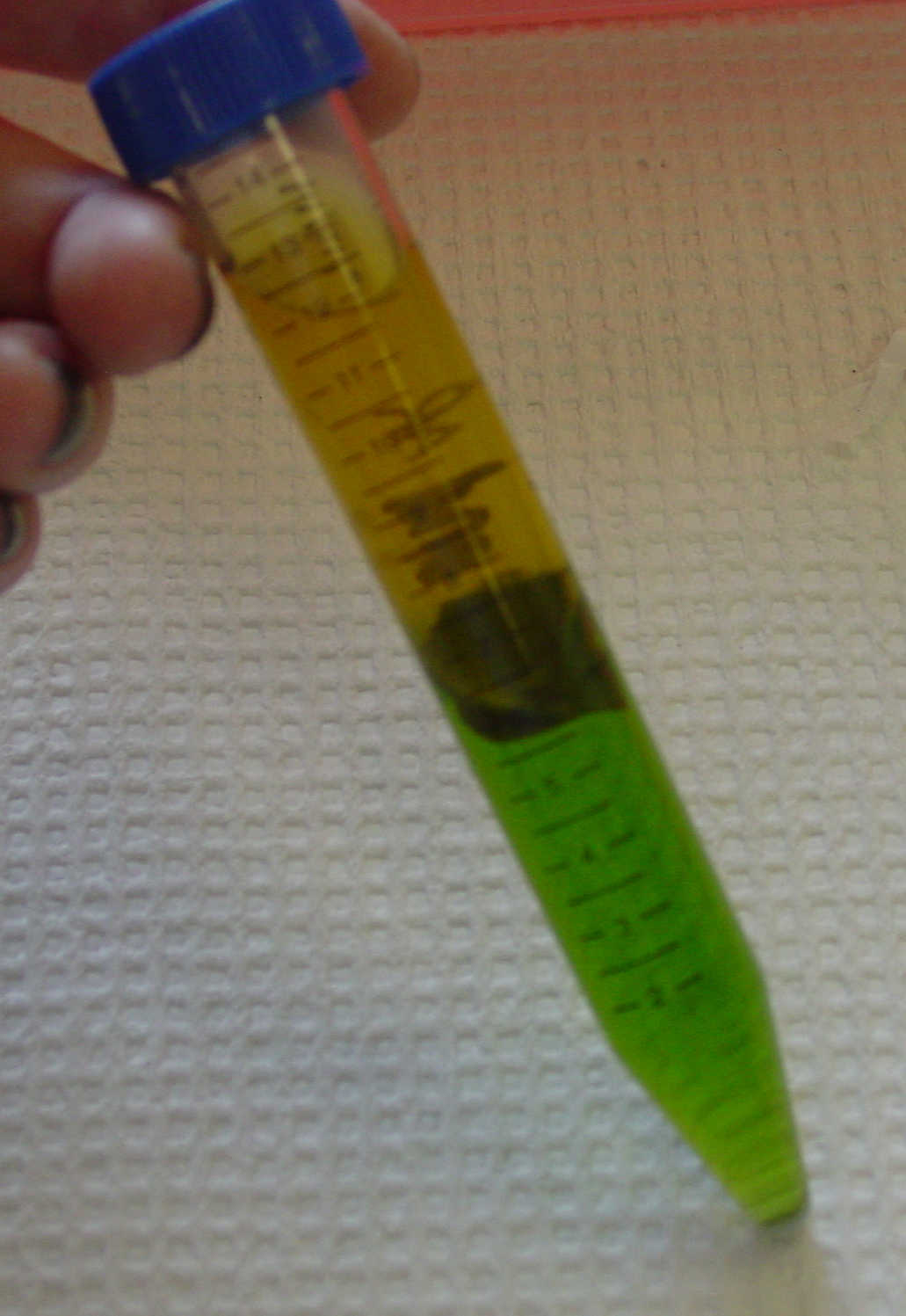
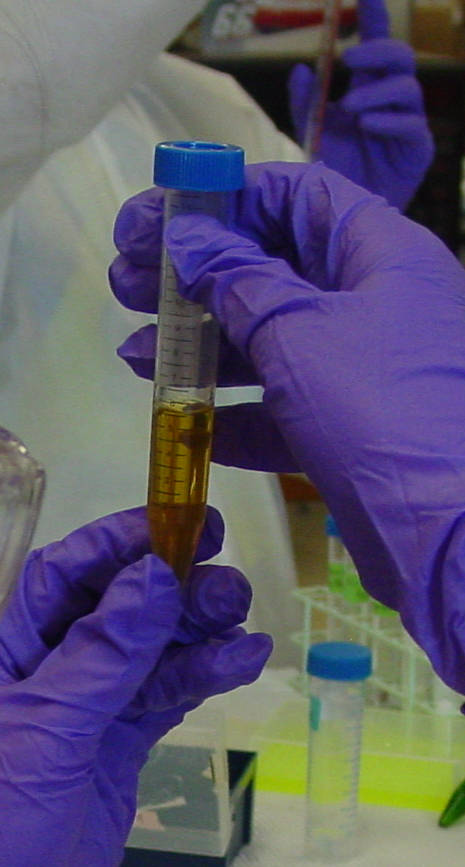
After mixing with chloroform and spinning in a centrifuge, 3 layers can be seen - we want the top straw-colored layer.
The top layer has our DNA, we pipette it into a new tube and add isopropanol to precipitate the DNA.
You can see the DNA come out of solution (white stringy stuff towards bottom of the tube).
Tuesday, June 17 th, 2014. The B3 Summer Science Camp Blog
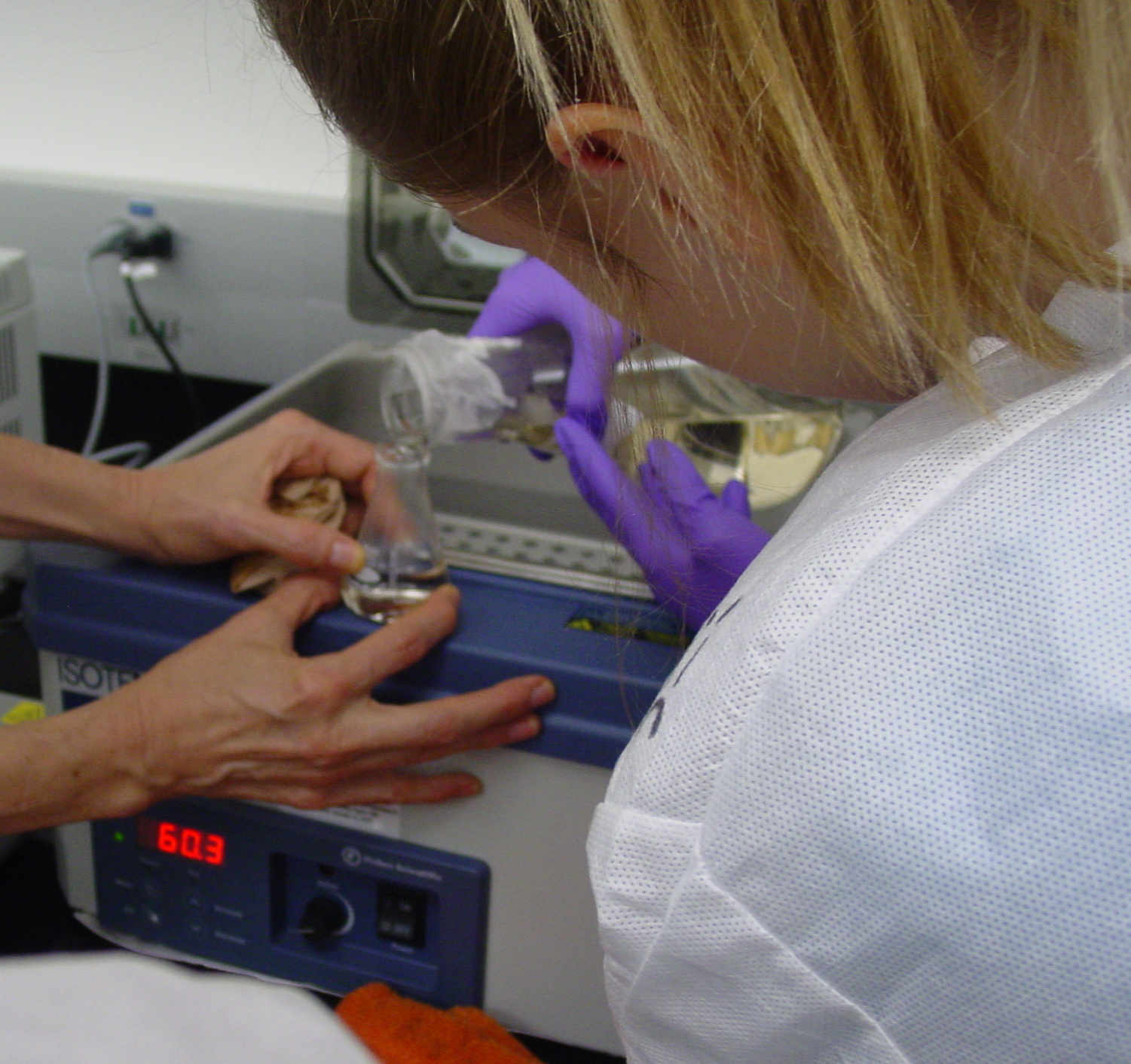
The real work began... with a Chestnut DNA extraction lab that required lots of patience and precision. Our main goal was to extract
the nucleic acid from leaf tissue. We started by grinding the leaf up in a chilled mortar with dry ice (to keep the sample fresh.) We added
beta-mercaptoethanol, as well as a few other chemicals to help break the sample down and pull the other parts that make up the leaf away from
what we really want... DNA strands. It sounds simple, right? WRONG. It took a lot of time to safely accomplish our goals. We had to have the
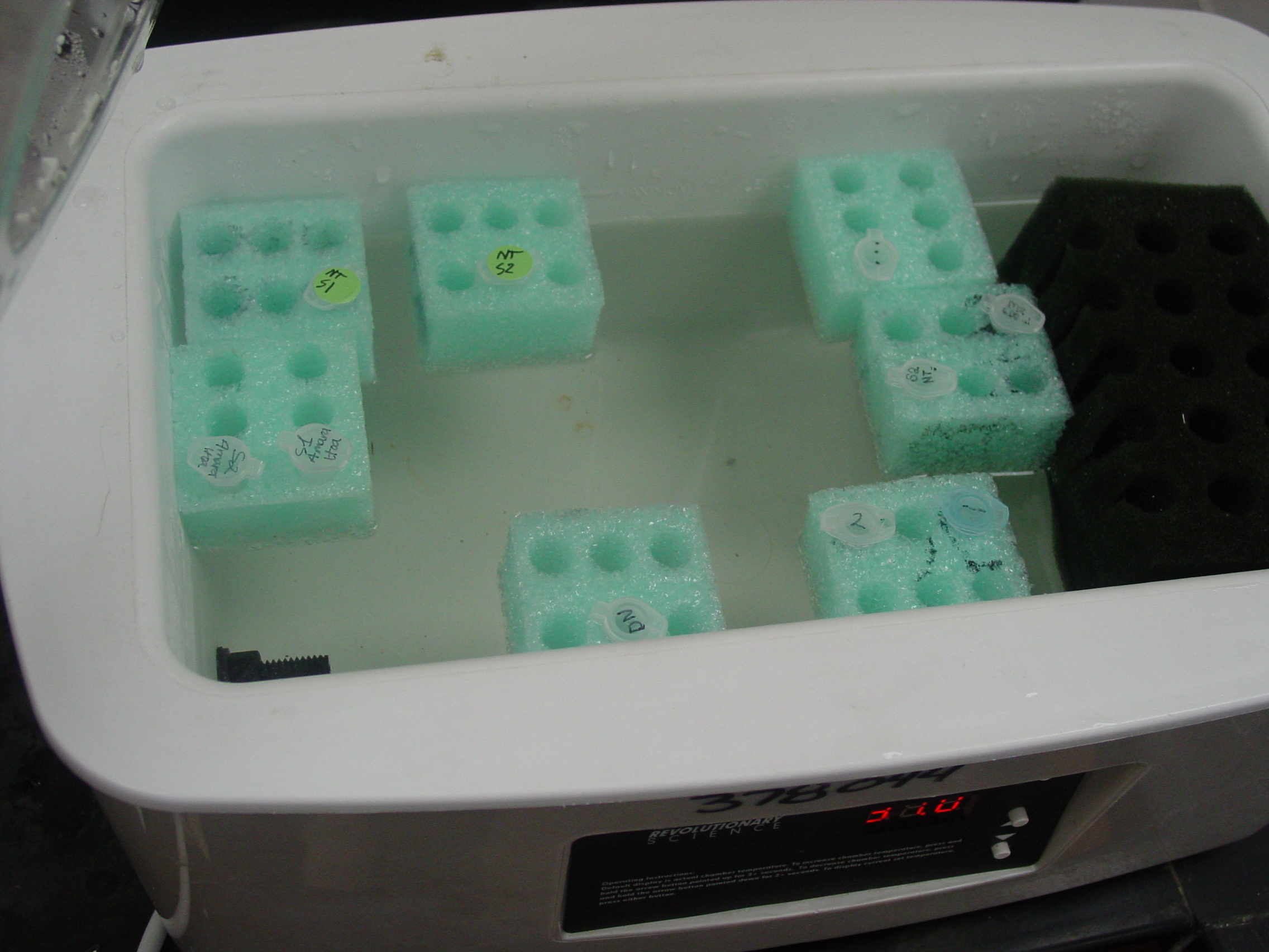
patience to sit with our samples in the water bath and the centrifuge (which basically spun them together.) Before we parted today, we left
our samples to sit over-night in 99% isopropanol, which will allow us to spool out the DNA in the morning. Of course, once again, there will
be many steps we have to follow, but we'll see how that goes tomorrow! ----- Annabel Richards
June 18 th
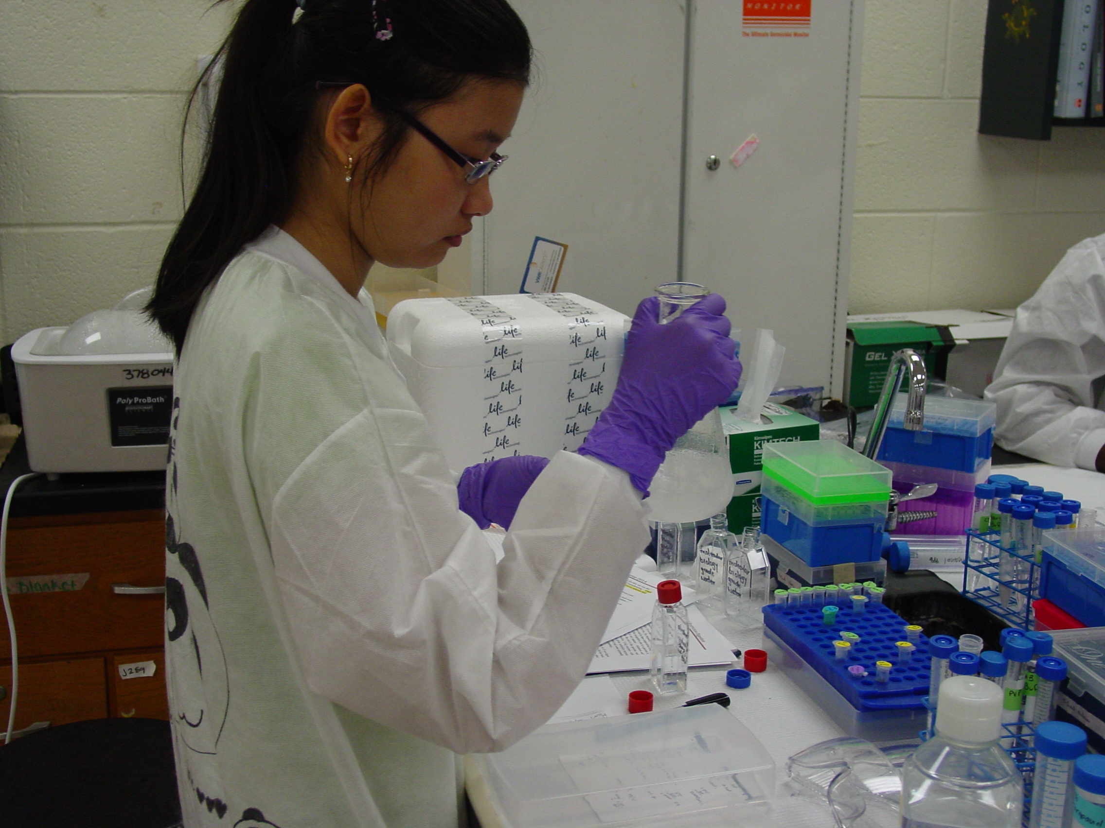
Continued DNA Purification - removing more carbohydrate and protein from the Leaf Nucleic Acid
Plants have a lot of carbohydrate and it takes a lot of work to remove all of it.
After spinning out our 'DNA' (which is really both RNA and DNA mixed with carbohydrate and protein) we re-dissolved it.
Then we went through several more purification steps.
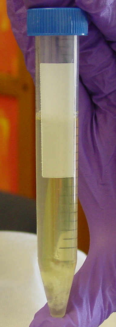
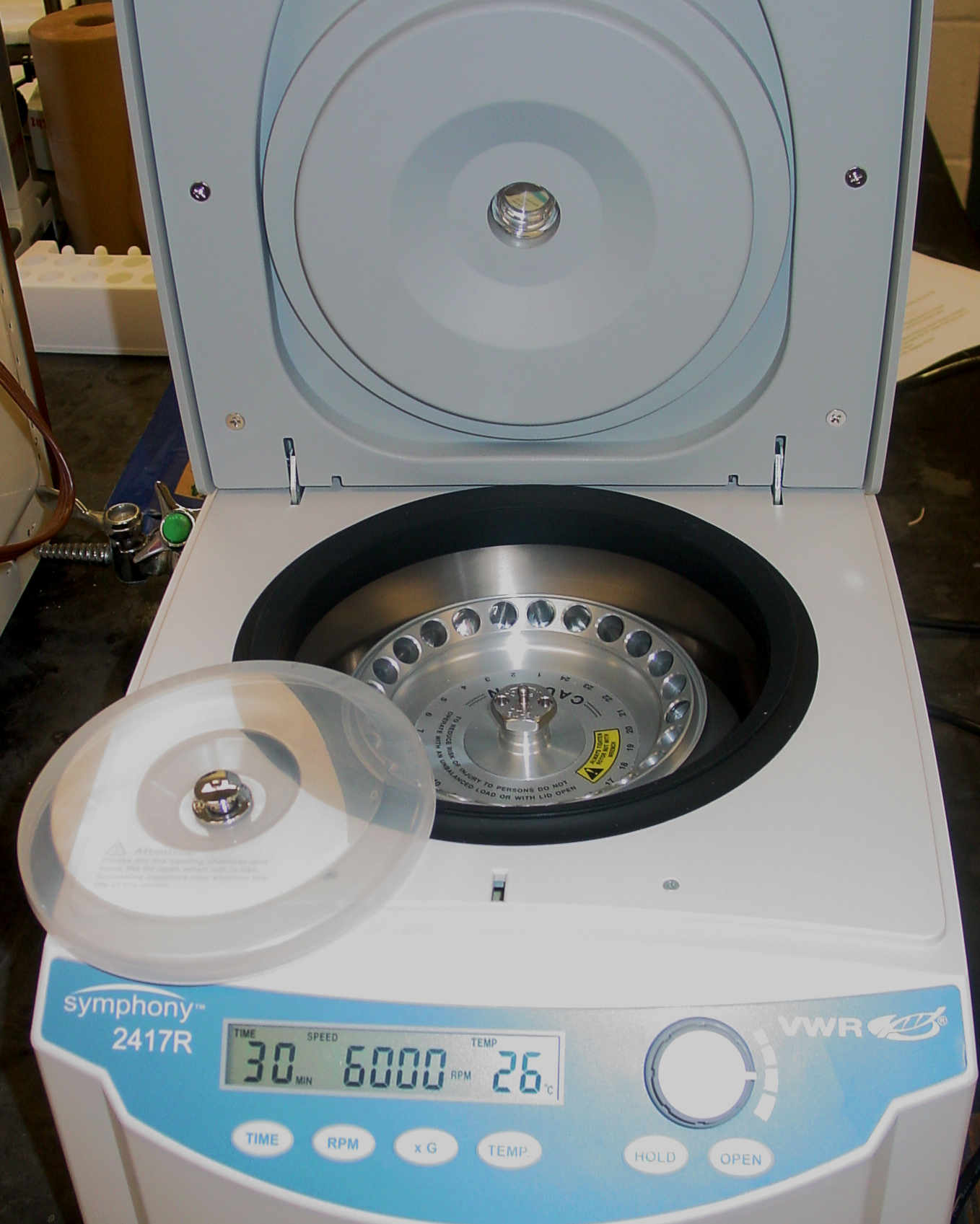
After centrifuging the tubes for an hour, the DNA/RNA is in a tightly packed pellet in the bottom of the tube.
The liquid can be removed with a pipette, and then the sample can be re-dissolved in buffer.
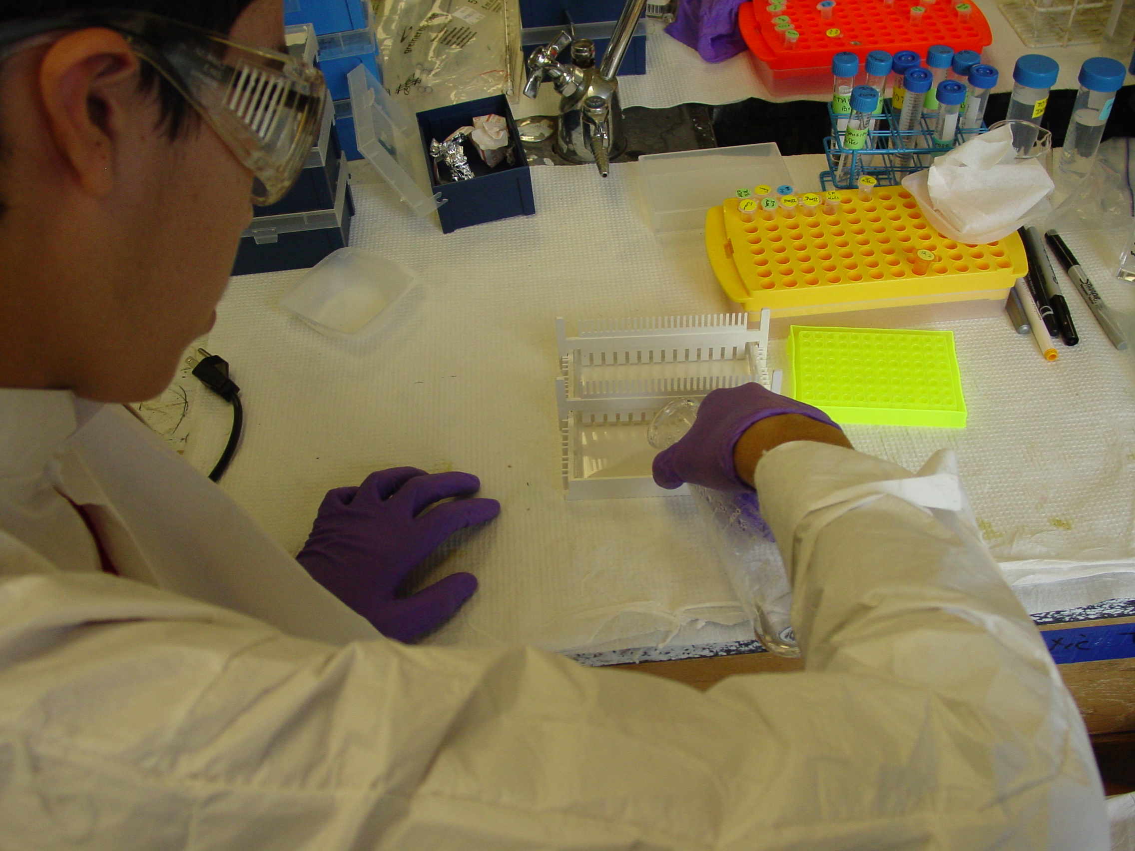
A vortexer mixes up the samples very quickly. Then the mixture can be moved to a smaller tube.
The smaller tubes are spun (centrifuged) very fast to collect the cleaned-up DNA/RNA in the bottom of the tube.
The DNA/RNA is re-dissolved by being shaken in buffer overnight on a vortexer with a home-made tube attachment (thanks, Jayden).
Wednesday, June 18 th, 2014. The B3 Summer Science Camp Blog
We received our samples this morning with a lovely pellet at the bottom (which is a good thing!) Our work began immediately, as we had to
drain the isopropanol and drain all the liquid from inside the test tube to isolate the pellet. We then added resolubilization buffer and put in
a shaker to fully solubilize the nucleic acid. The main goal was to break up the pellet to release any trapped DNA. After we took it off the shaker,
we stuck the samples in the microfuge to spin. We basically repeated the same steps a second time, with the exception of added chloroform. We removed
the top layer (which wasn't a pellet, but it was definitely what we wanted) and transferred it to a fresh 1.5ml tube. Once again, we added isopropanol,
spun them in the microfuge, and were left with a pellet. We left the samples in the lab with the pellet and 200 microliters of TE buffer. I apologize
if I've lost you within the steps; it's MUCH more exciting to see everything actually happen. While we've been stuck on the chemistry part of it all,
work more linked to biology begins tomorrow (as we grow closer and closer to FINALLY obtaining our DNA.) ----- Annabel Richards
June 19 th
Even More DNA Purification - This time we really mean the DNA part
It is time to remove the RNA and the last bits of protein.
- this is done by adding enzymes, proteins with activity in breaking down RNA (RNAaseA) and protein (Proteinase K - which also degrades itself).
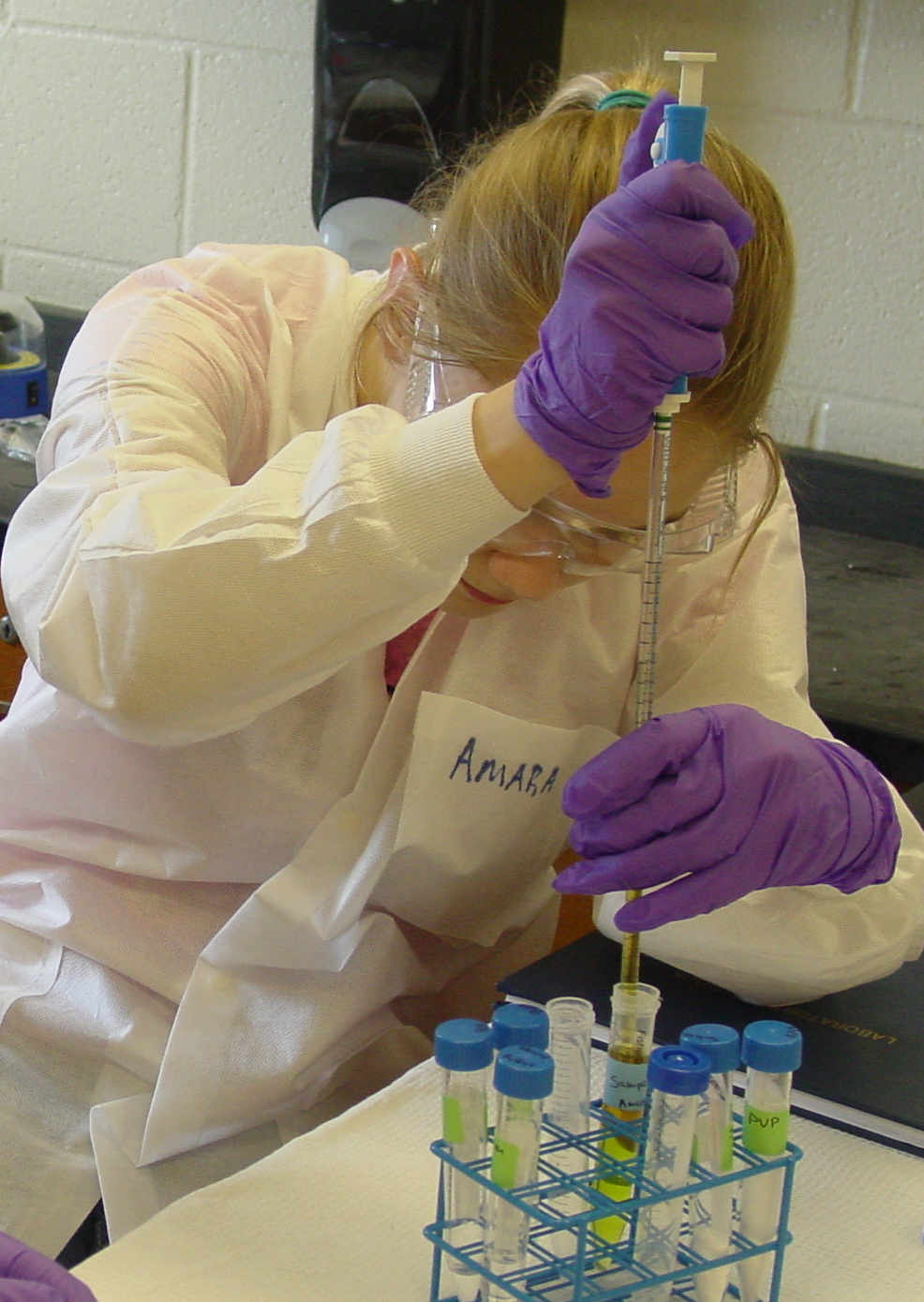
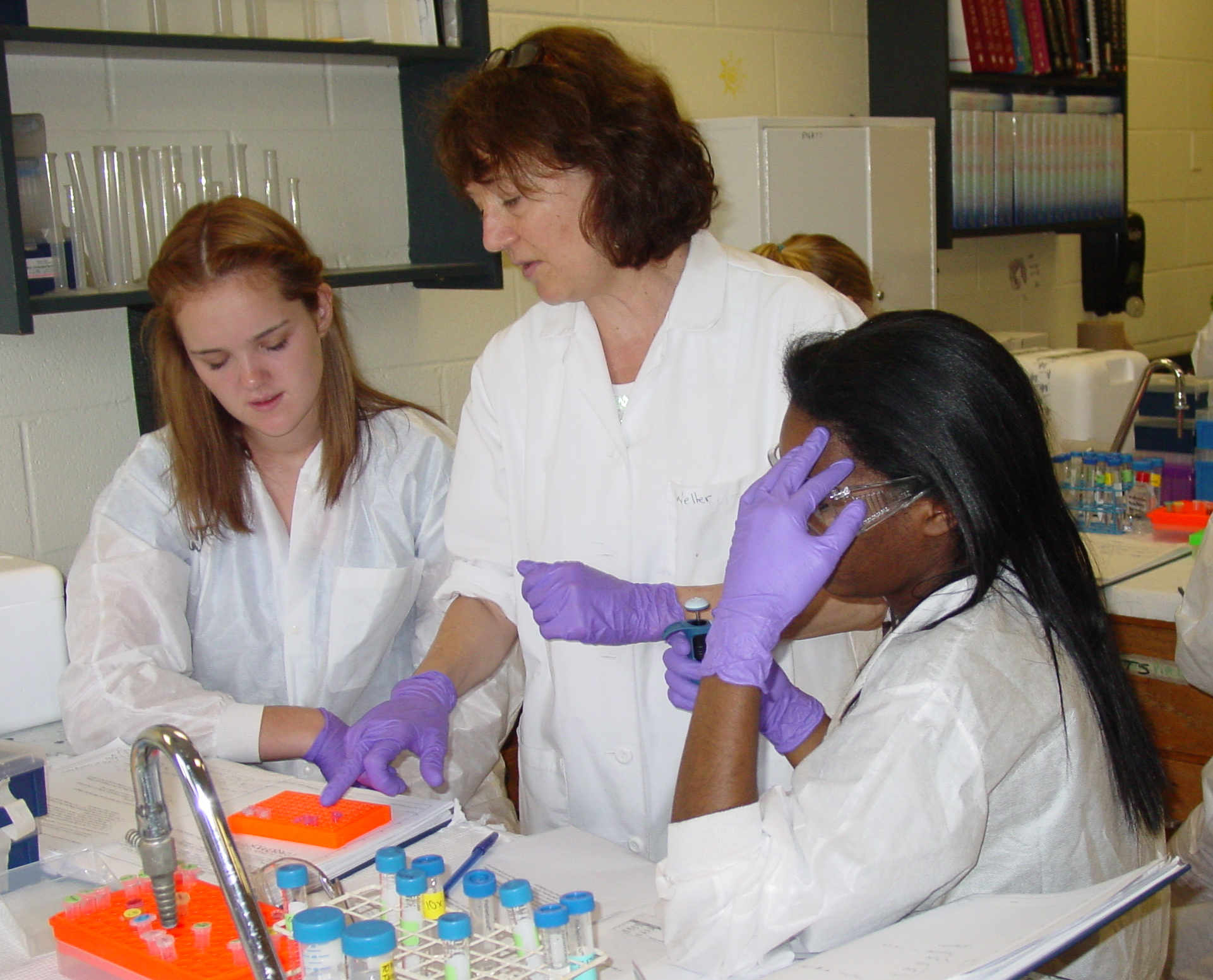
There is a lot of pipetting and centrifuging and waiting and then repeating.
We used the waterbath, 3 types of centrifuges and a lot of small tubes today.
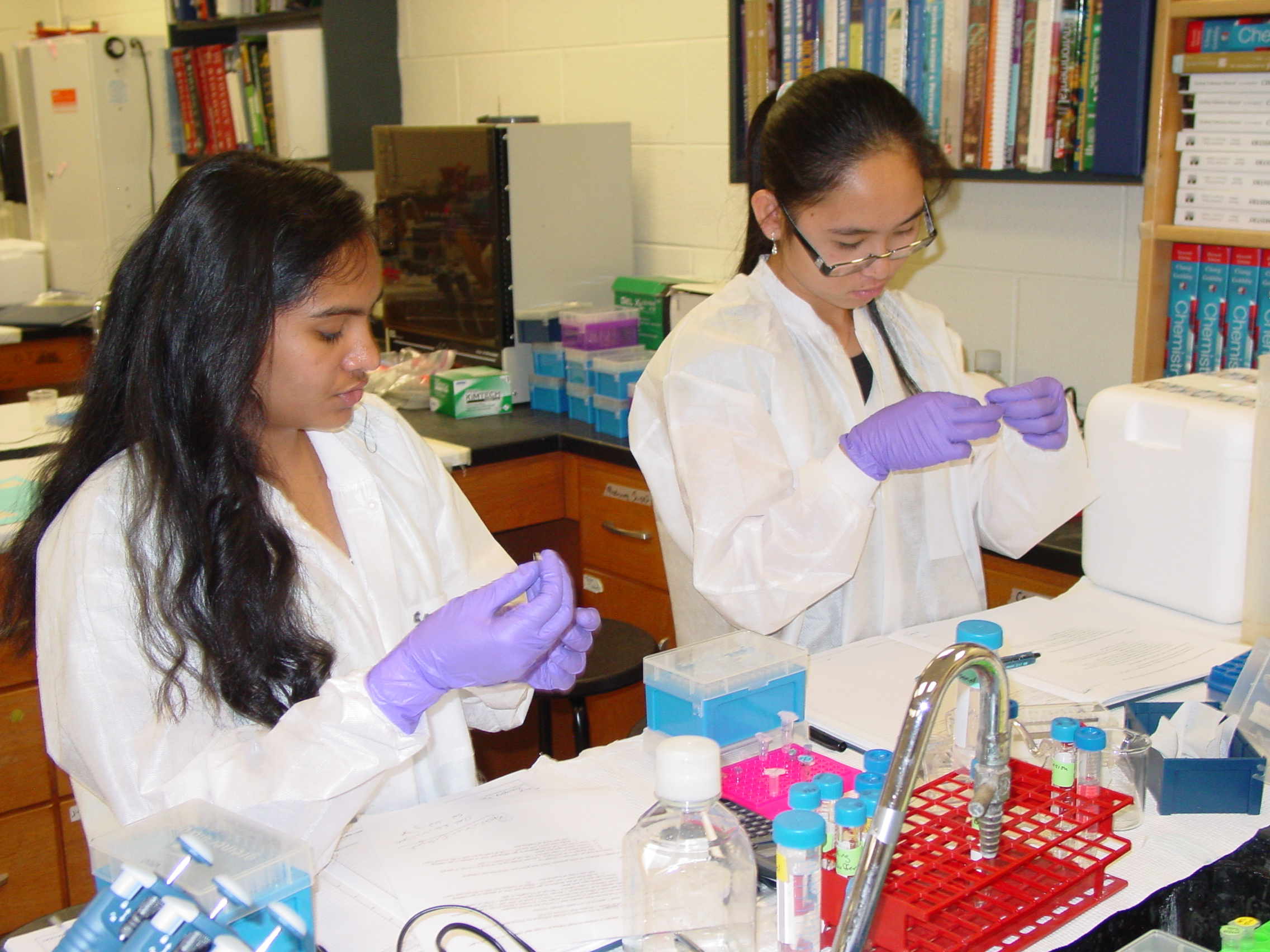
We did a lot of pipetting
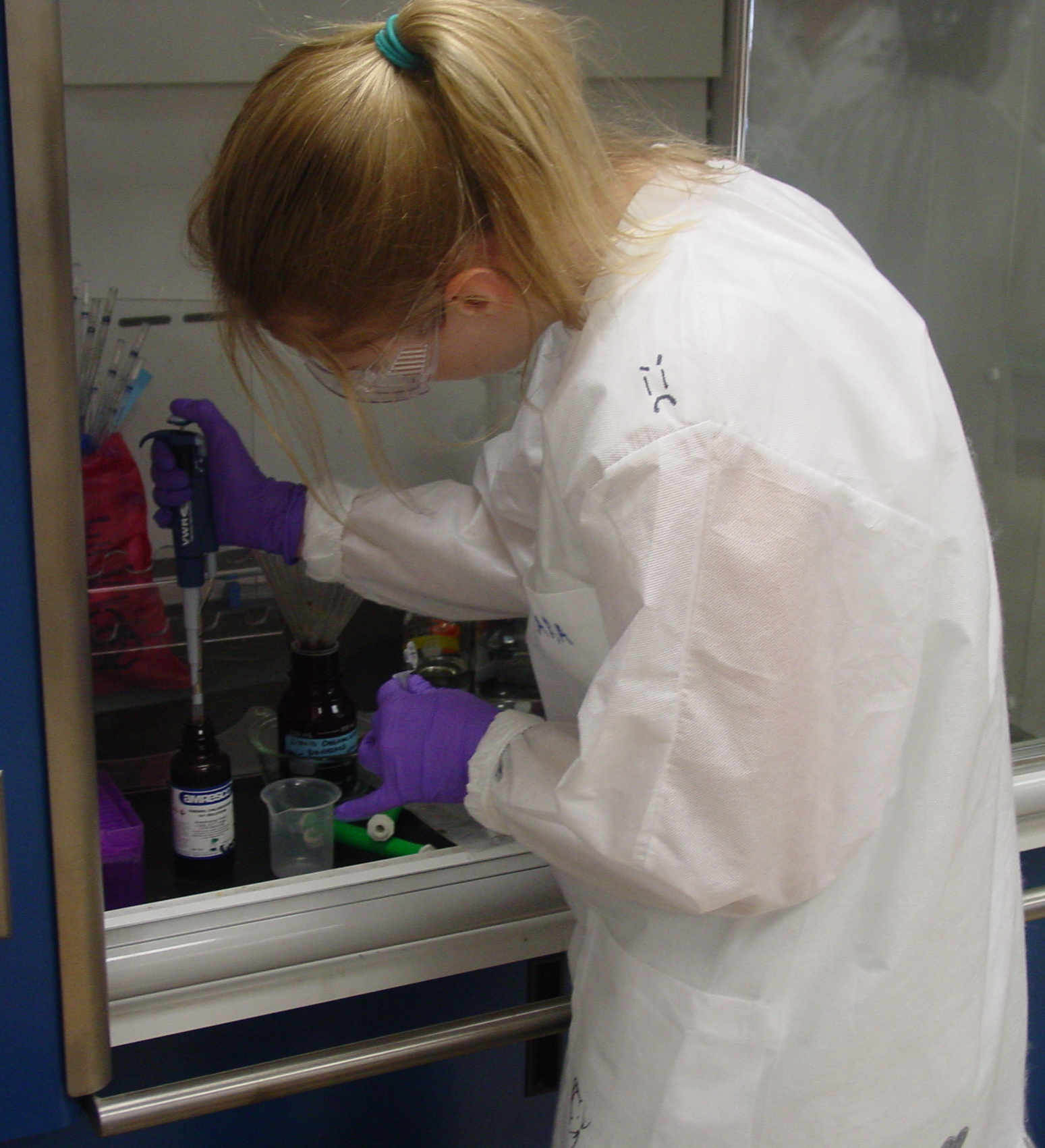
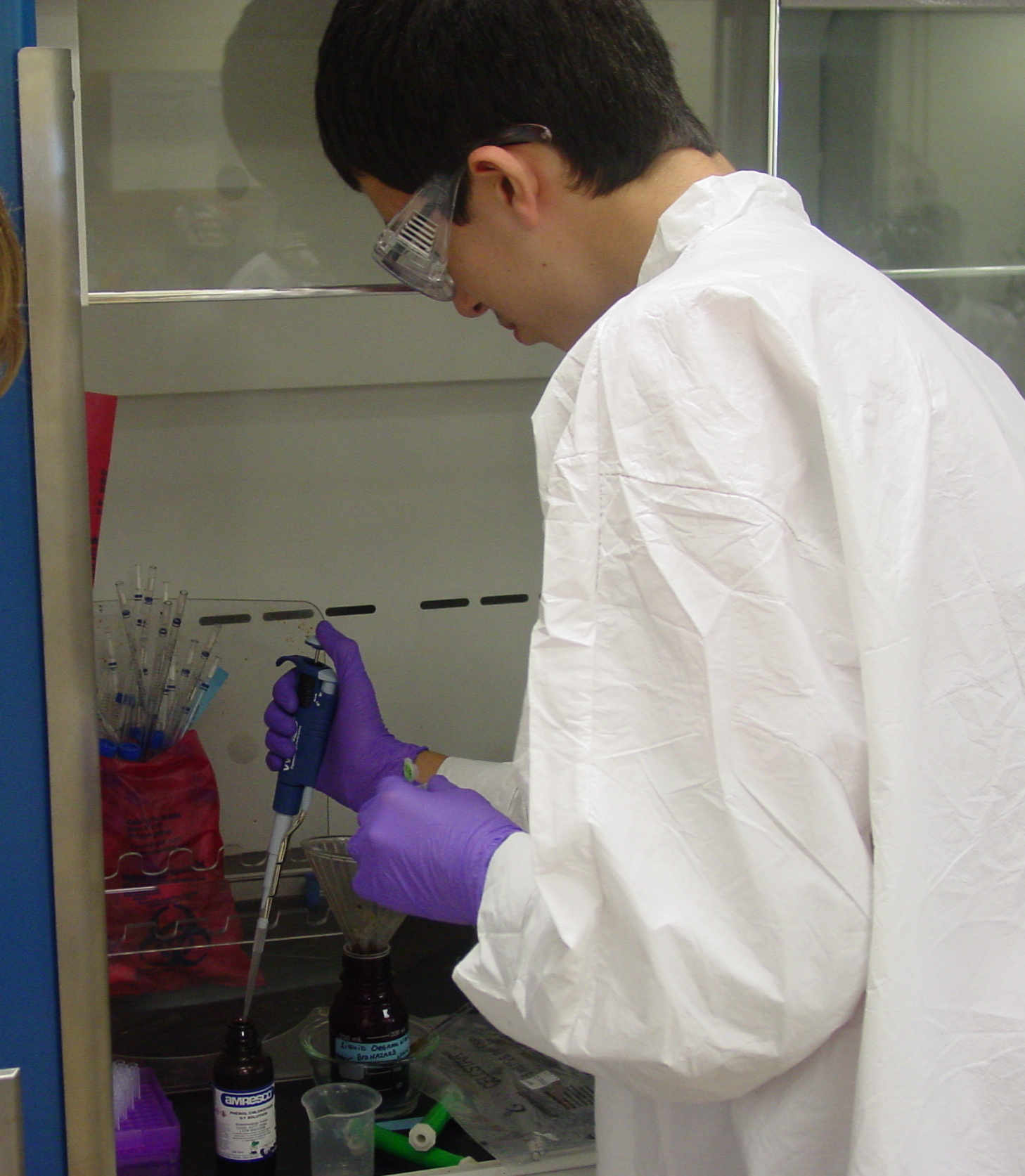
We added Phenol-Chloroform to remove all of the protein, wearing protective clothing and using the hood.
Did we mention we did a lot of pipetting?
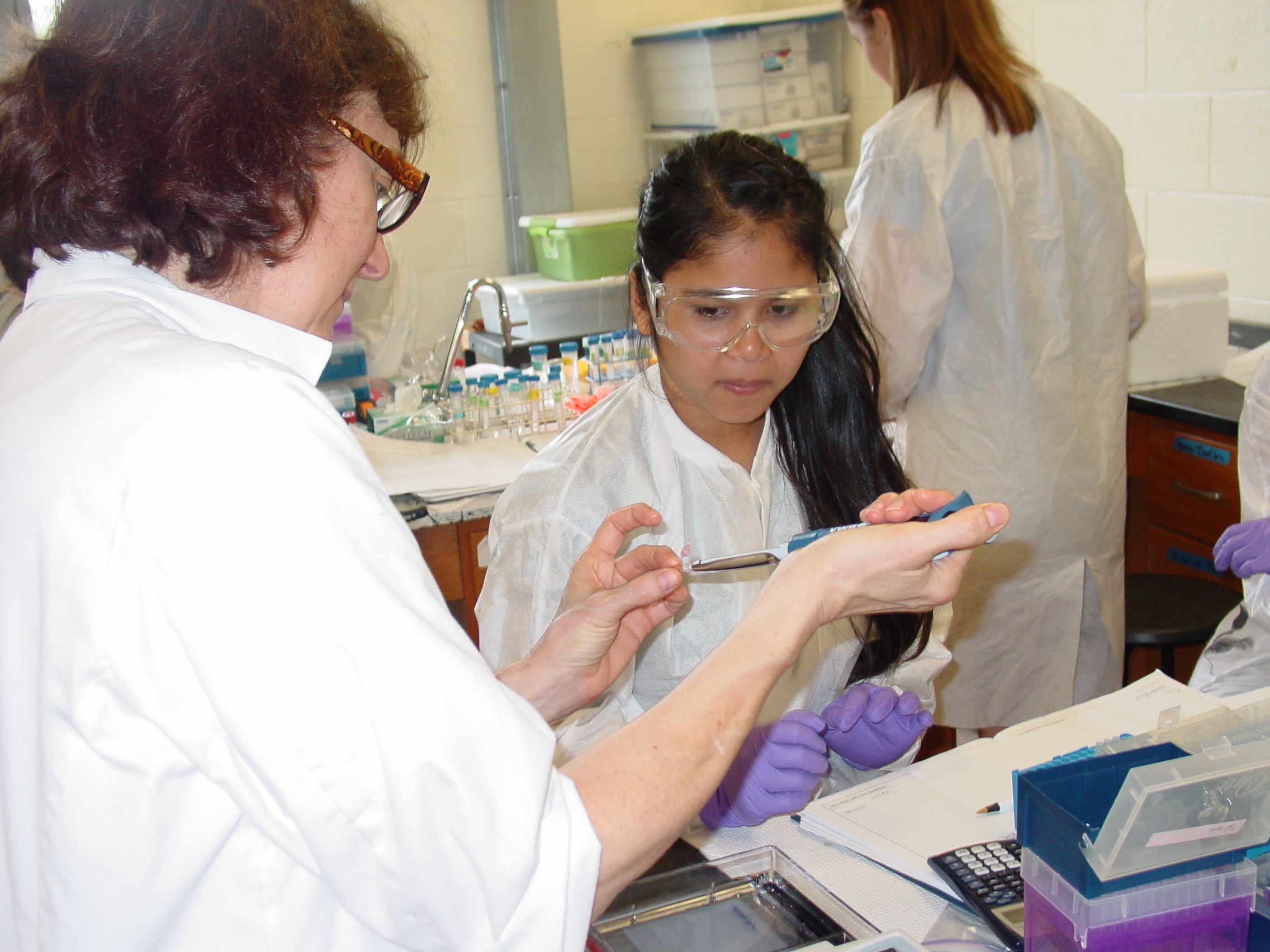
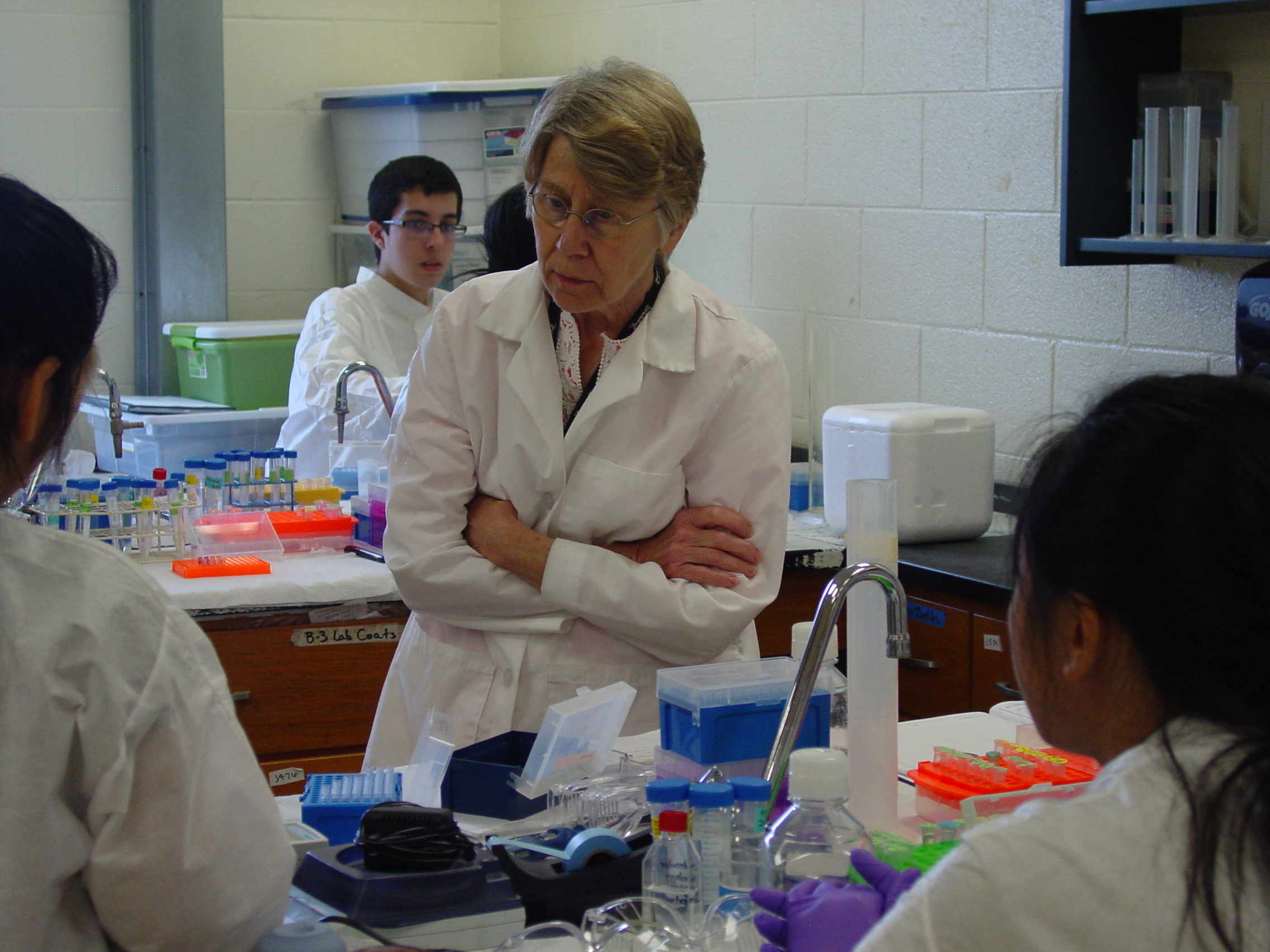
Samples have to be balanced properly in a centrifuge, Ms. Putnam double-checked our tube placement.
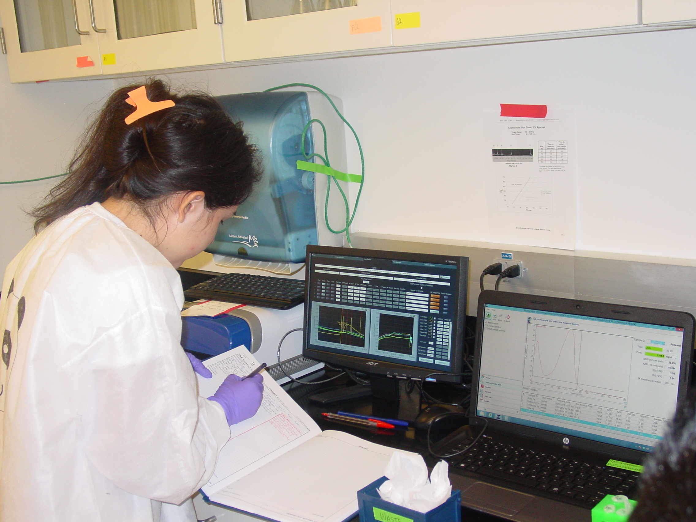
We also checked out the spectrophotometer using some samples Jayden prepared, so we will be ready tomorrow.
Once the samples were spun we had to remove the liquid and find the small white pellet on the bottom of the tube.
And there was a lot of waiting - it was really nice to get to the end and see we all had some DNA.
Thursday, June 19 th, 2014. The B3 Summer Science Camp Blog
We spent today doing all that was left of purifying our DNA, and removing the remaining contaminants of our sample. This consisted of more
spins in the microfuge, and the addition of chemicals such as RNase A, Proteinase K, Phenol-Chloroform, Isopropanol, and 3 M NaOAc. There were several
moments when we had to wait for our samples to spin in the microfuge or sit in the water bath, so instead of waiting, we had two very important
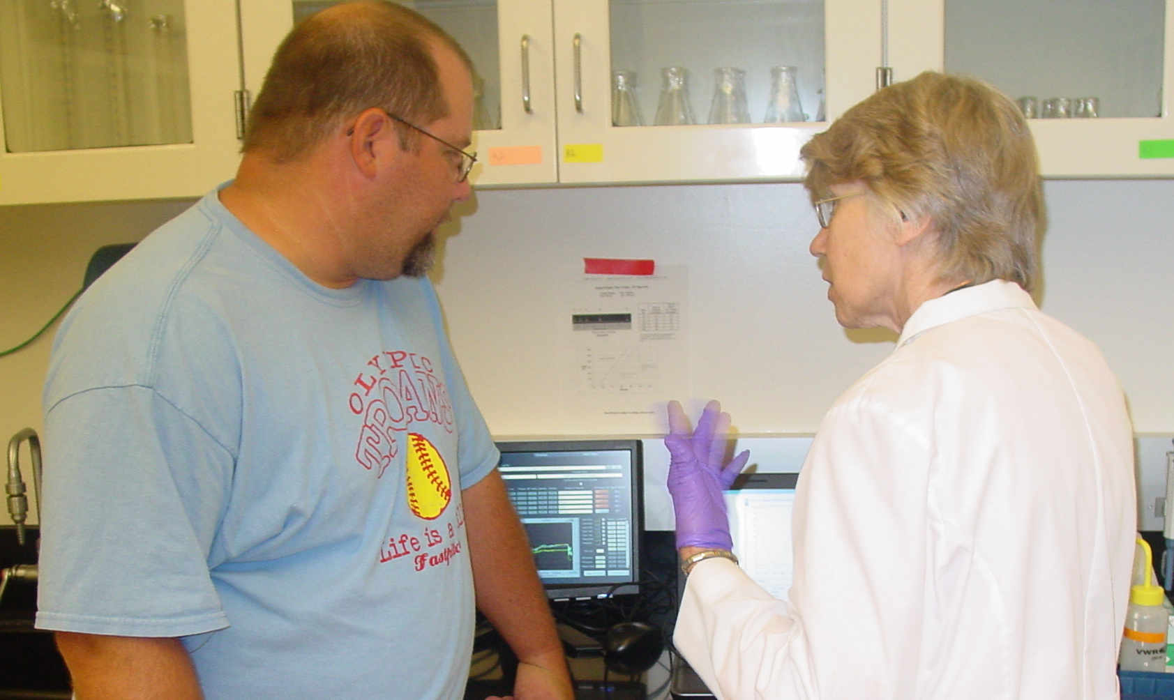
introduction discussions about things we were using in the near future. The first was a lovely lesson about spectrophotometers, taught by Ms. Smith.
To sum things up, a spectrophotometer shines a light on the sample in solution. The molecules (or in our case, the DNA) interact with the light.
The light wavelength chosen, or the data it reports back to you, is based on maximum absorbance. This is important, because it will allow us to
verify there is nothing left contaminating our DNA samples. If the ratios reported back are much lower than what they are supposed to be, then
we know there is still something in our sample. We haven't used this lovely machine yet, but based on it's description, it's going to save us a
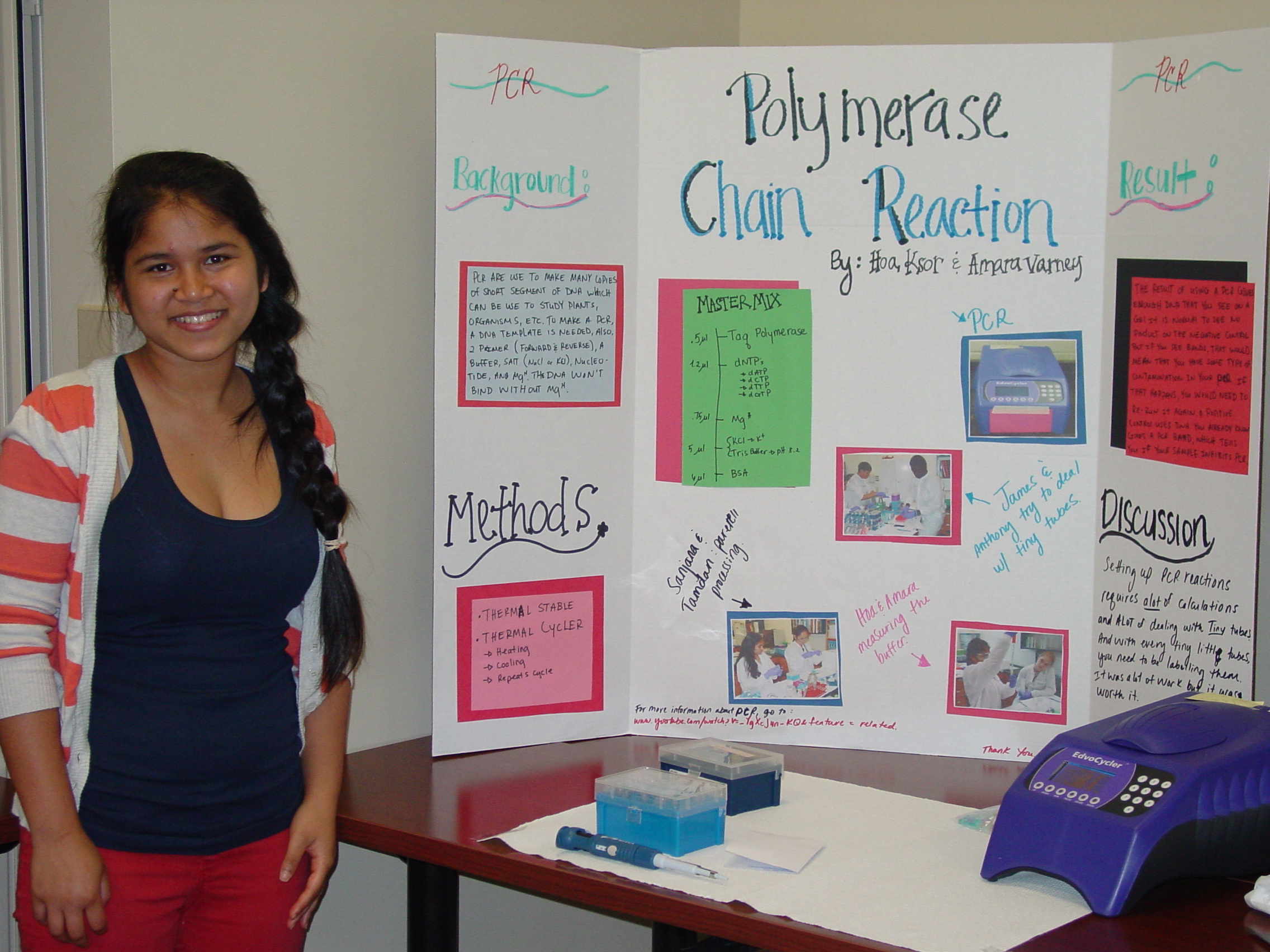
lot of time and possible irritation. The second discussion was about polymerase chain reactions. To sum things up once again, a polymerase chain reaction
is a big fancy way of describing the copy machine for DNA. PCR reactions allow you to replicate specific sections of your DNA until you have a good amount
of copies to work with. This will also help us a lot, because we will actually be able to see our DNA strands. ----- Annabel Richards
June 20 th
Field Trip to Dr. Weller's Lab at UNC Charlotte
We are going to do some of our work in a research lab, and also visit some of the other labs.
- The schedule for the day was quite ambitious:
First, use a sophisticated spectrophotometer to determine how much DNA we had and check its purity .
Second, perform restriction digestions on 2 micrograms of our DNA, which will cut it into smaller pieces in a reproducible way.
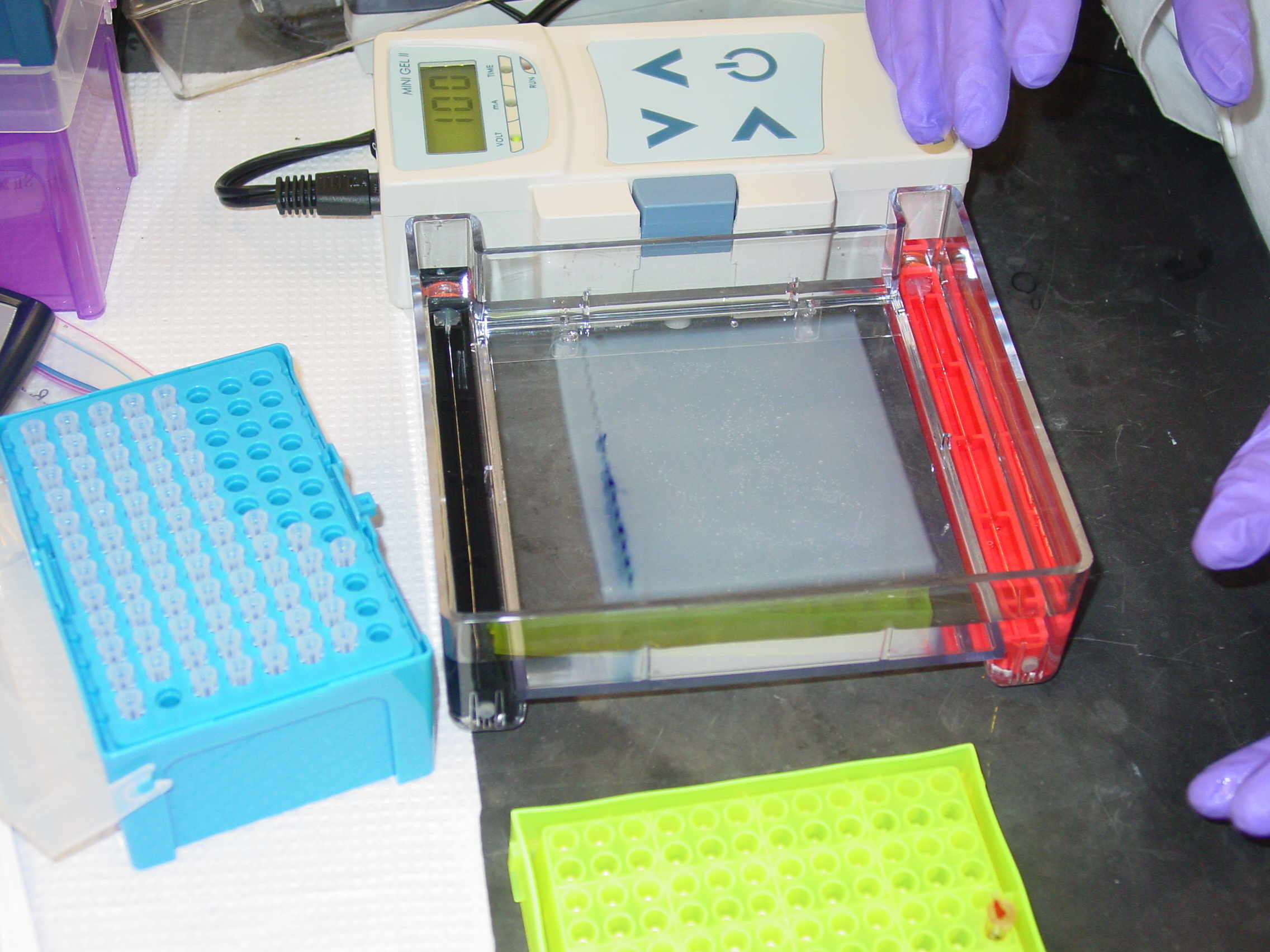
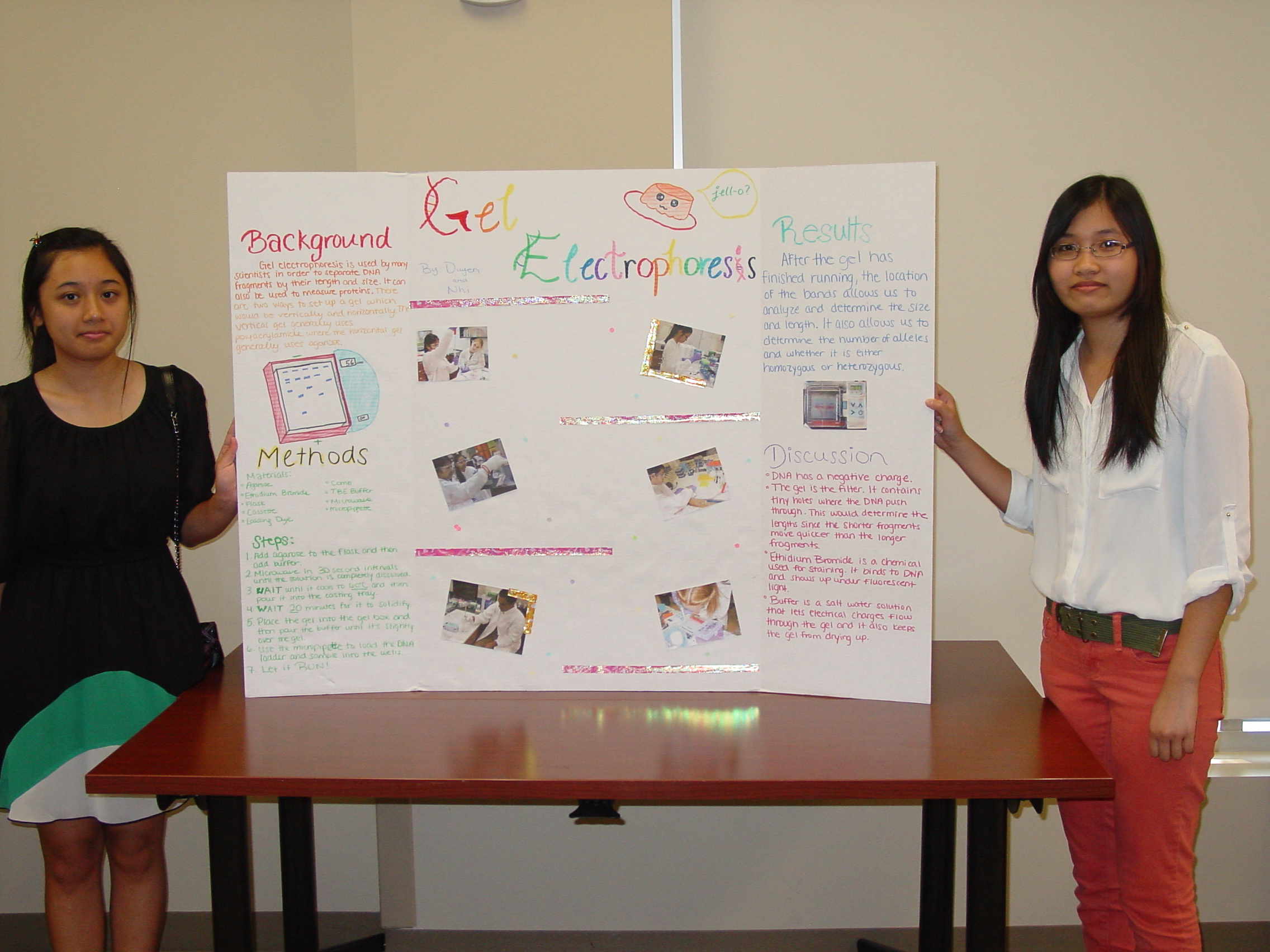
Third, carry our agarose gel electrophoresis on our DNA before and after the restriction digestion to check the amount and lengths visually.
Fourth, use magnetic beads in Pelyethylene Glycol to separate DNA by size for future uses.
Finally, tour the Bioinformatics and Genomics labs to see what other technologies are used to carry out this type of research.
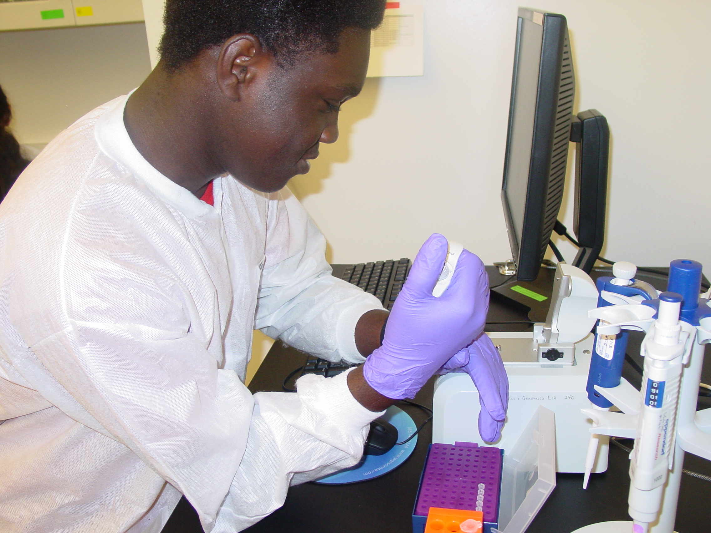
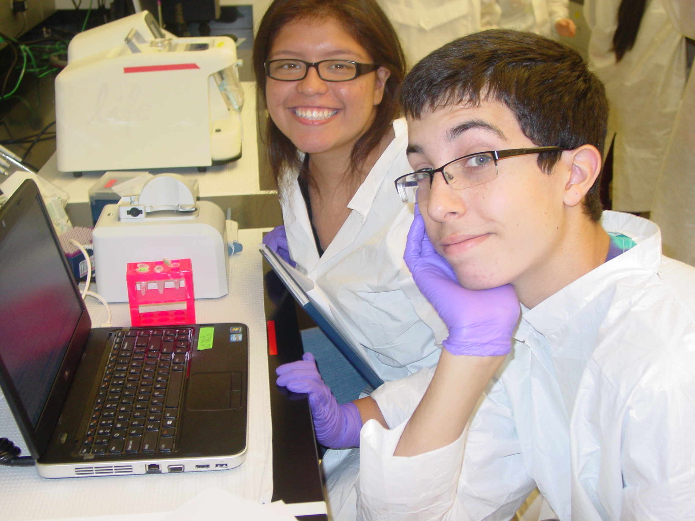
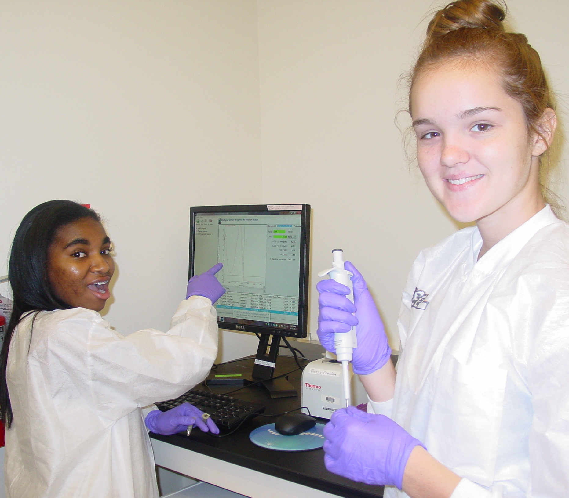
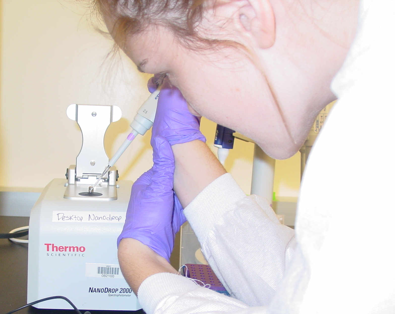
We had 3 Nanodrop UV-Vis (Ultraviolet-Visible) spectrophotometers for the class to use.
James Tripp and Nicole Doudgie and Annabel
Getting the droplet right on the pedestal was important. Then we had to interpret both the shape of the absorbance curve and the concentration the spectrophotometer reported.
Careful Sample Placement Maria examines the absorption curve Amara checking calculations with Dr. Weller
Setting up Restriction Digestion requires a specific order for adding the solutions. Once that was done we poured agarose gels.
Duyen setting up the steps Ms Smith explaining the tasks to Jeremy Collecting agarose for the gel
Unfortunately, right after lunch a personal emergency required that the students leave early, so some of the tasks will be completed next Monday.
Students who participate in the Fall Semester Saturday Science club will re-visit the University Research labs several times.
Friday, June 20 th, 2014. The B3 Summer Science Camp Blog
Today we took a field trip up to UNCC, and were able to utilize the more efficient spectrophotometers in her lab. After we collected our data,
we had to do a lot of calculations for our restriction digestion protocol. Using the average concentration calculated by the spectrophotometer, we used
a specific equation to figure out how much water to add to our sample. We added 10x buffer, a specific enzyme, a specific amount of our sample, and water
to a fresh new tube. After that was all mixed together, we stuck them in an incubator for two hours. ----- Annabel Richards
Monday June 23 rd
Back in the Labs at Olympic High School
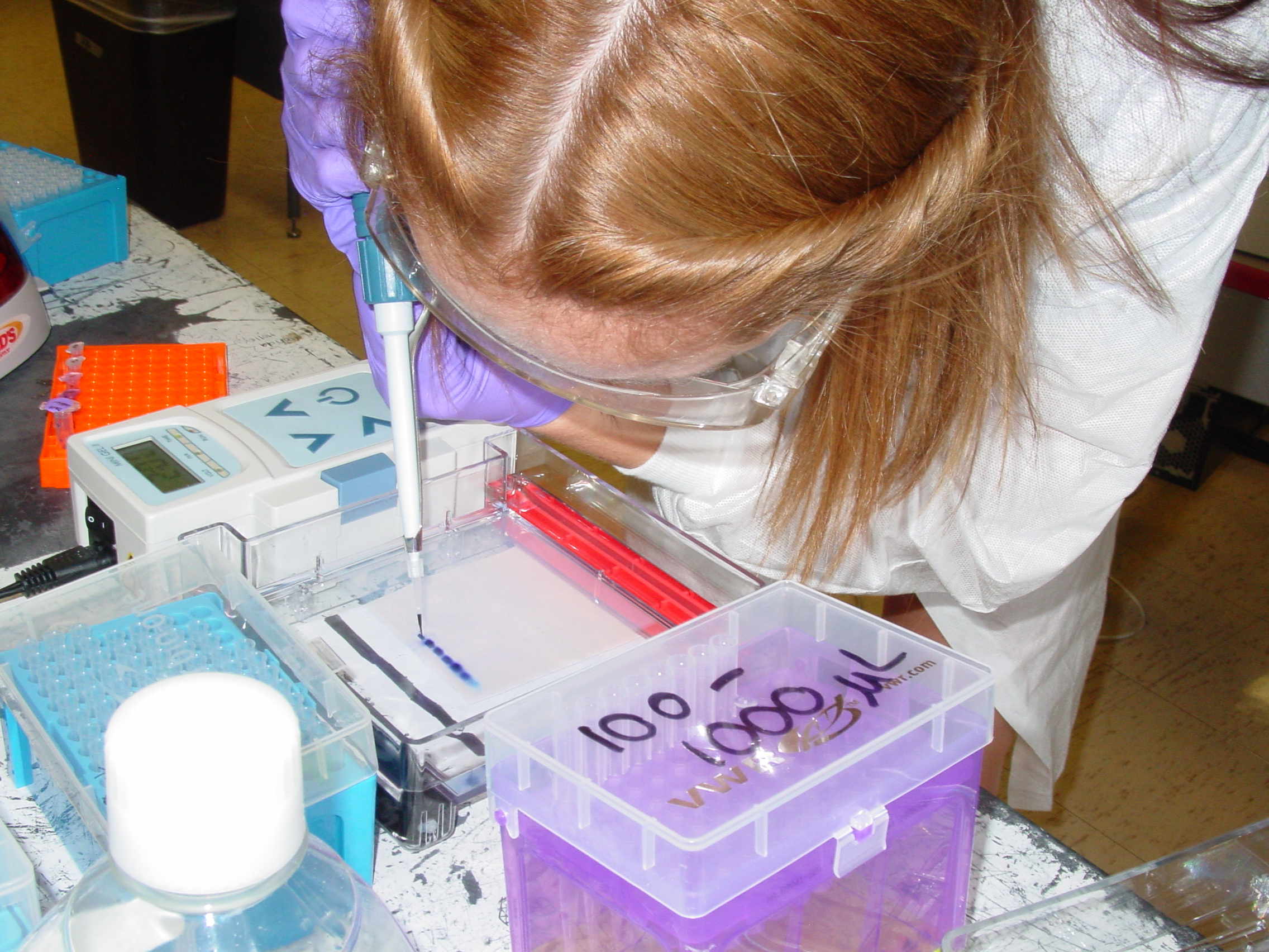
We will run some of our DNA on the agarose gels we poured on Friday that we did not get a chance to use.
We have to calculate how much sample to load and make a dilution.
Then we have to combine the sample with loading dye so it does not float away .
We have to include a DNA ladder so we know how big our DNA fragments are.
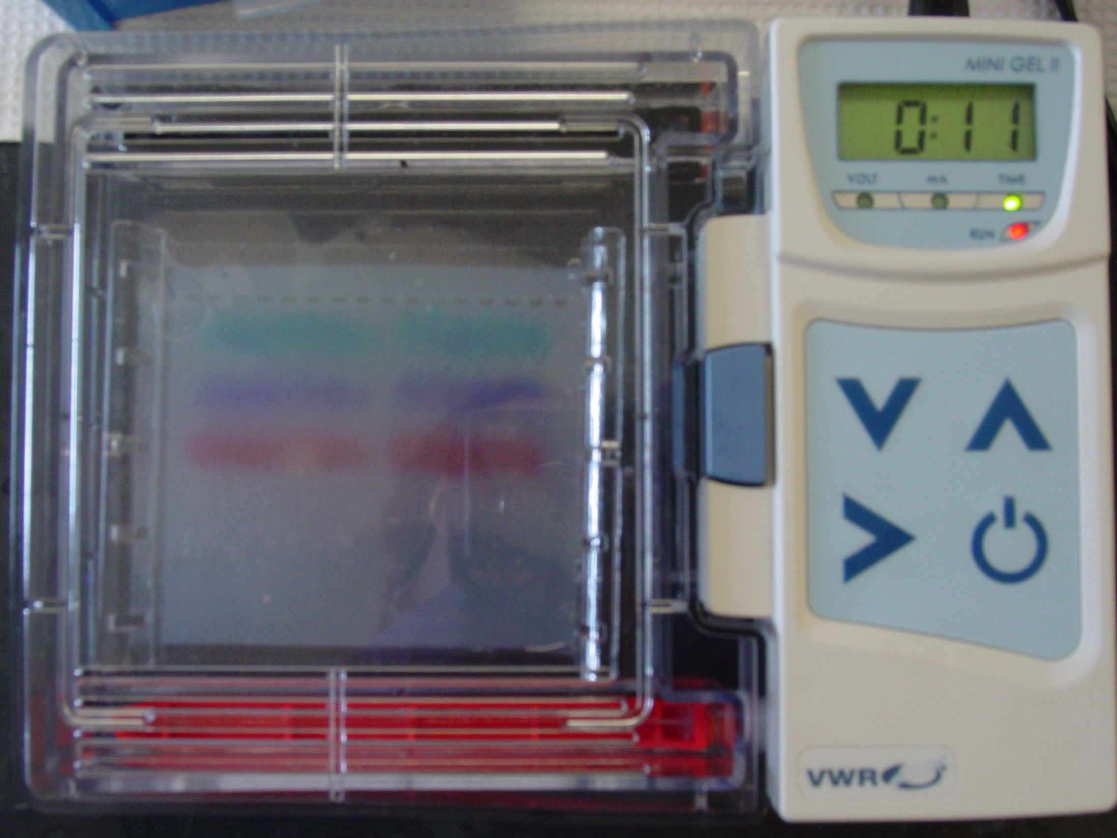
We included a dye that fluoresces when it is stacked with DNA, so we can visualize the DNA.
We also set up our first PCR reactions, which monitor the type of chloroplast in our trees.
Amara and Nhi Checking logic Maria asks Jayden for verification Dr.Weller, we are so confused! How to get rid of bubbles in a tip Minifuging the sample also works Super gel-loader This gel is ready to run Checking electrophoresis progress The dyes have migrated in the gel Setting up PCR reactions came next, and we had to do calculations for that too.
Maria and Willie with duelling calculators James and Anthony try to deal with tiny tubes Sanjana and Tamdan: parallel processing
June 24 th
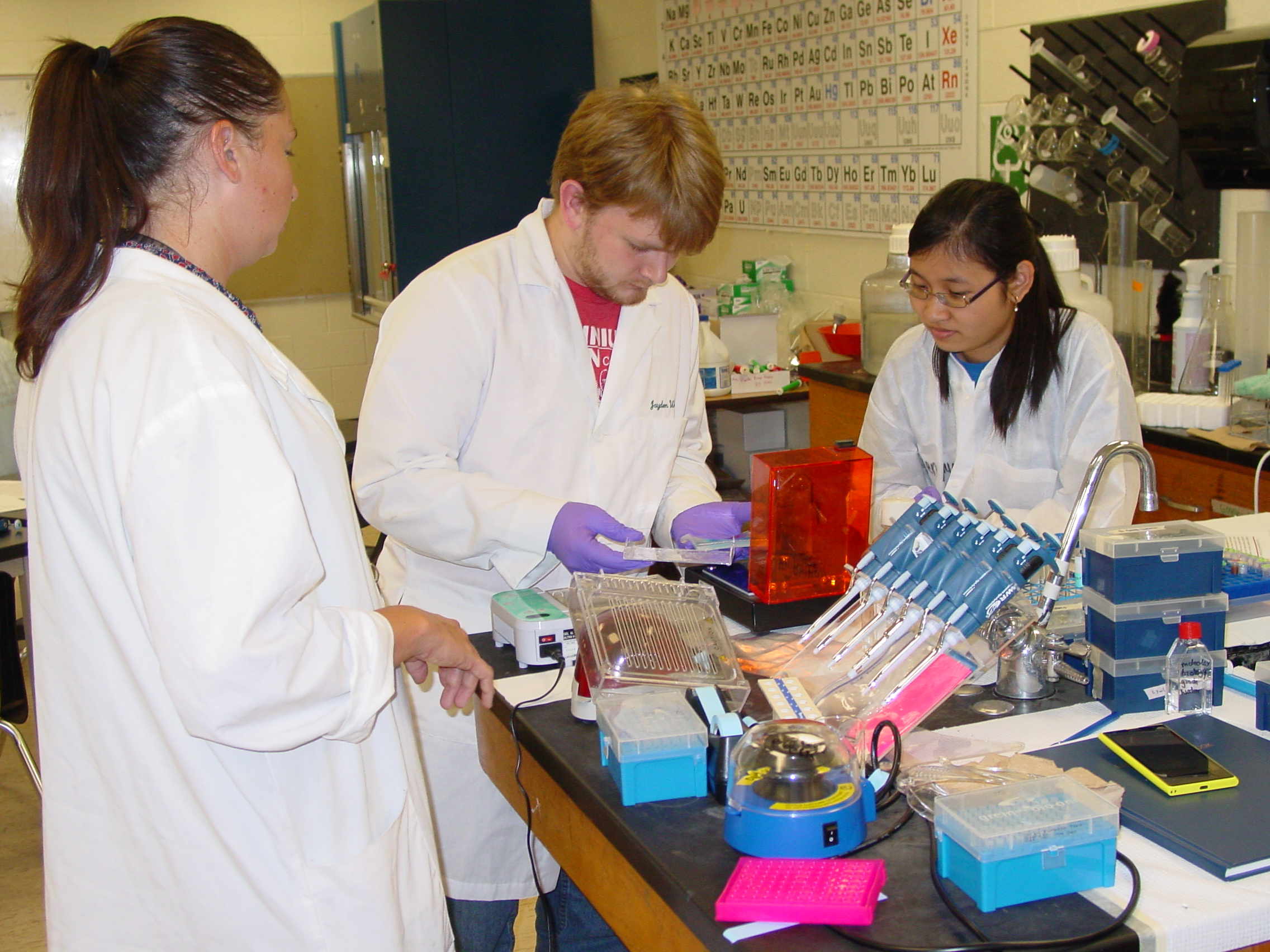
Pouring agarose gels and setting up second PCR reactions
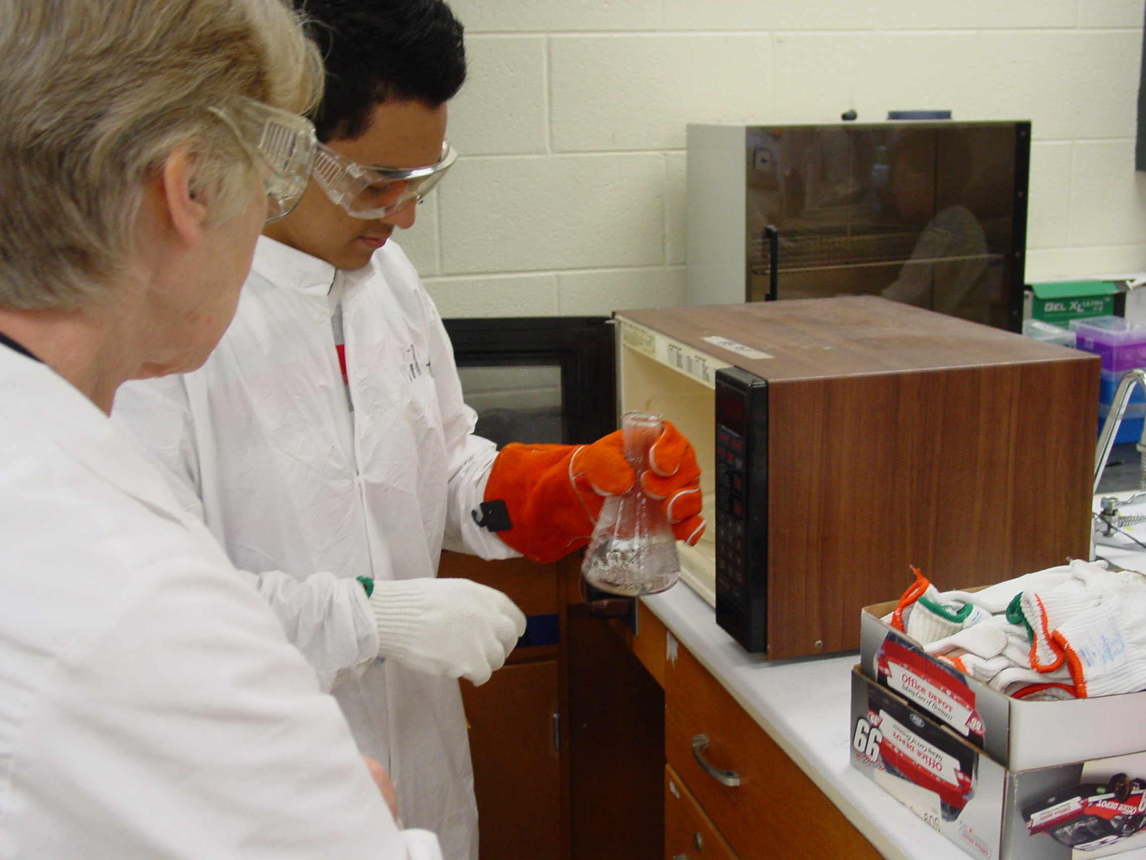
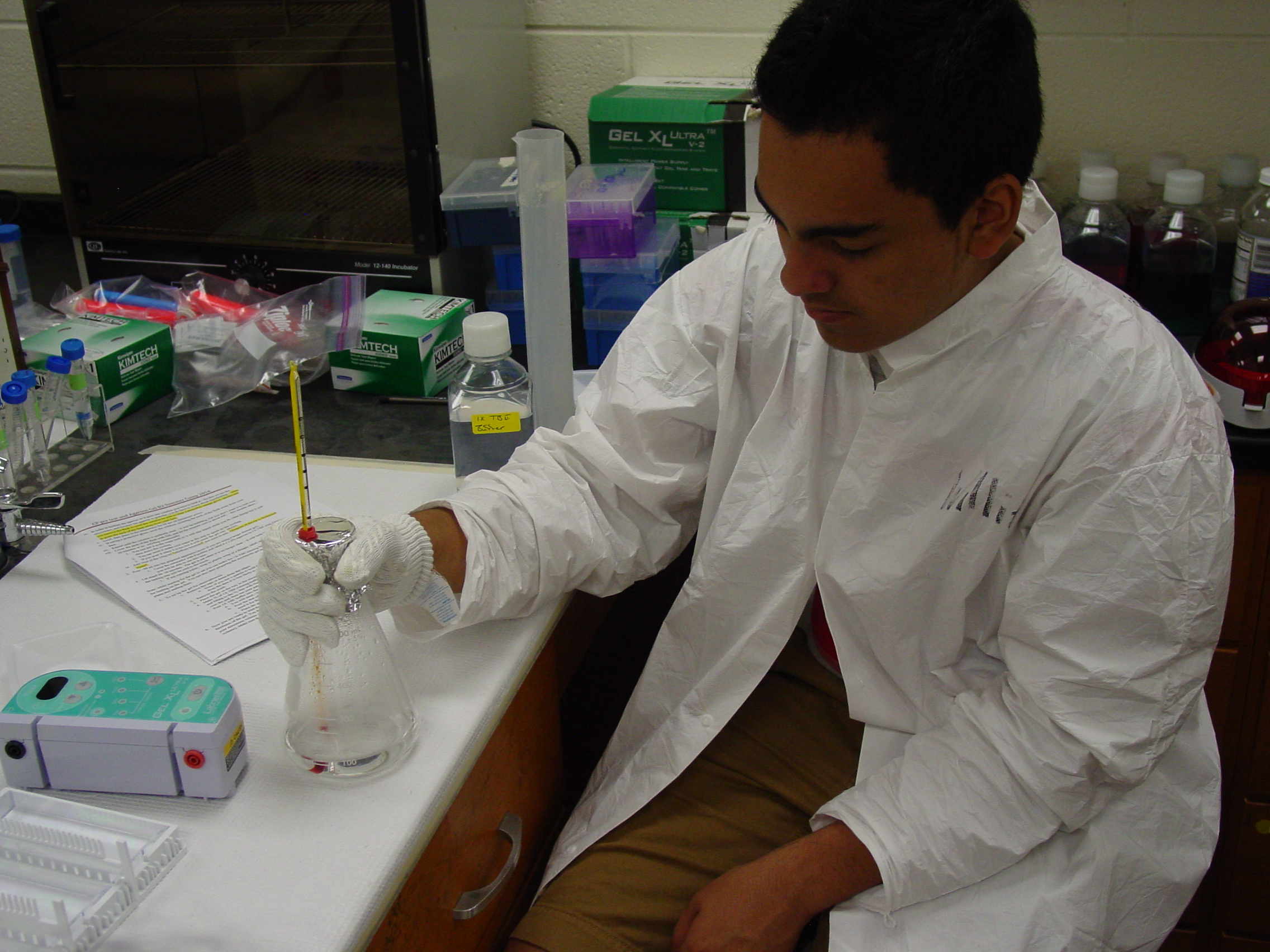
Today we learned to prepare the agarose for gels.
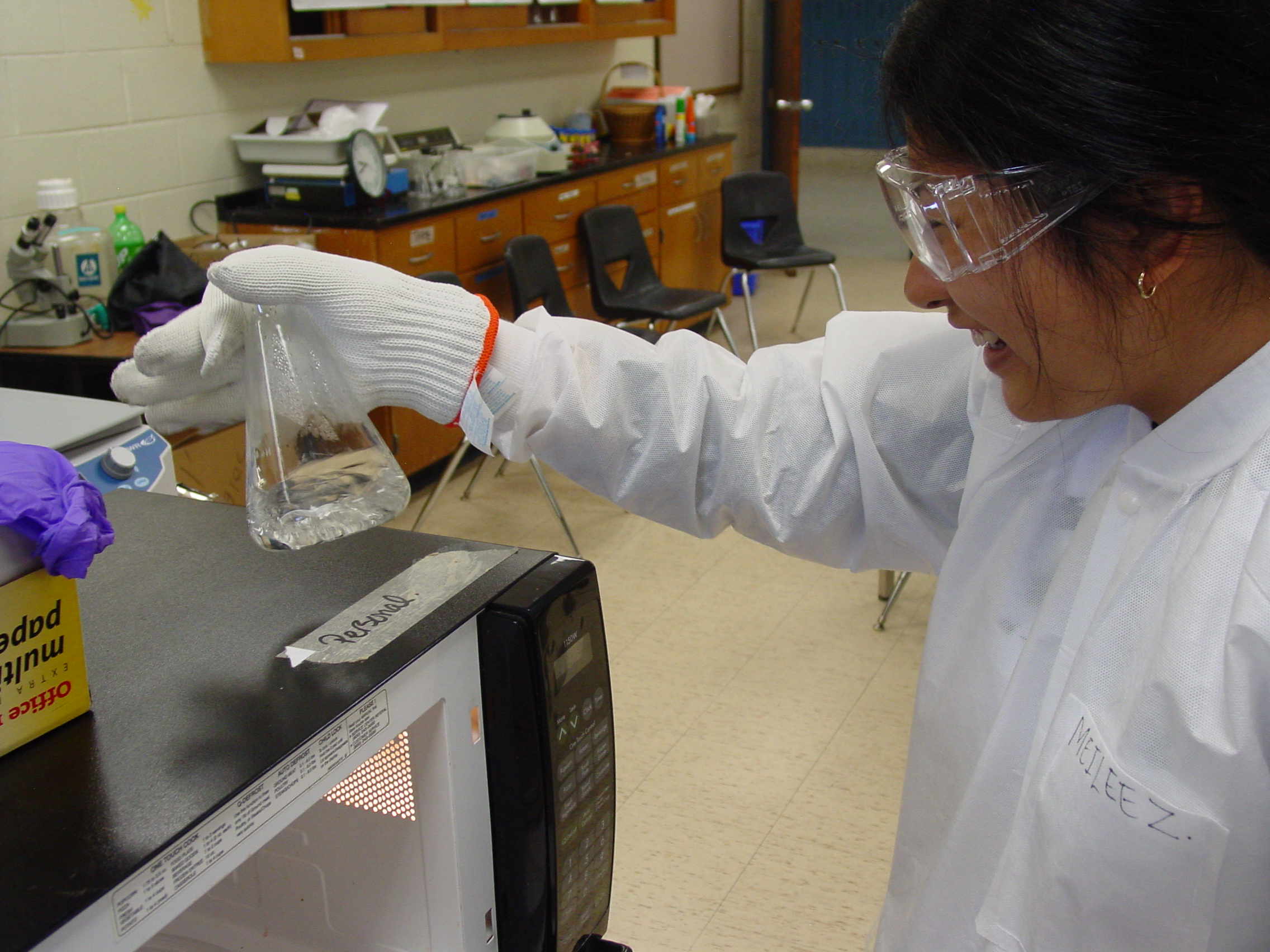
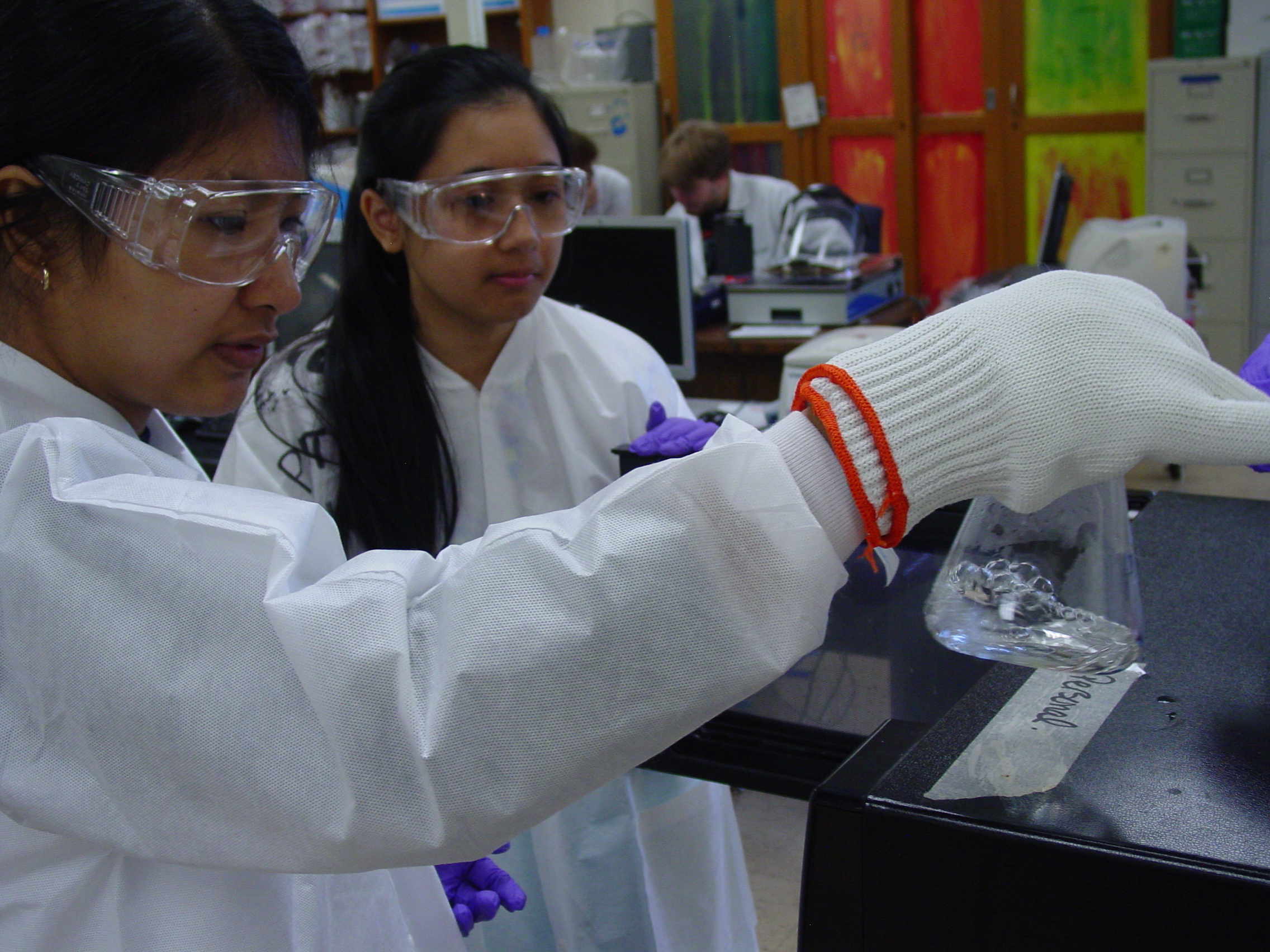
We learned proper safety precautions (gloves, lab coats, goggles).
We repeated and extended our PCR reactions to include additional markers. .
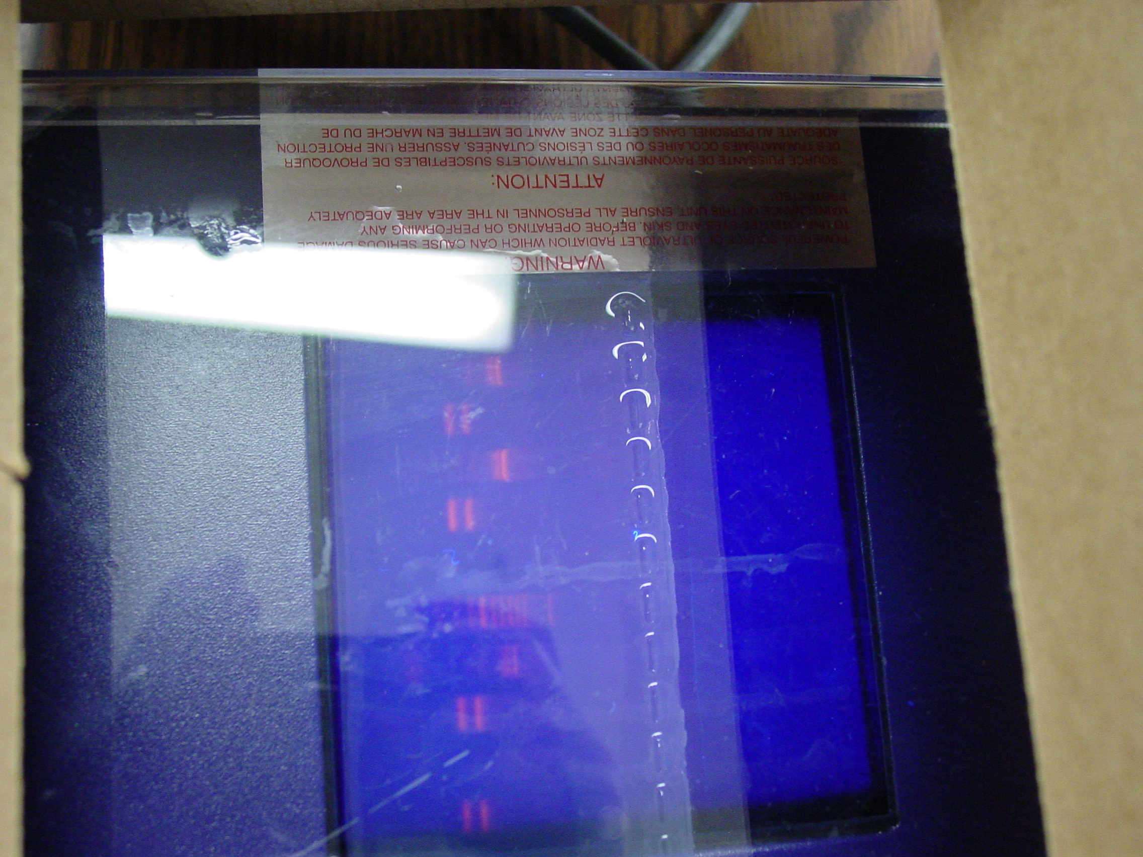
Amara and Hoa measure buffer Duyen lets the agarose hydrate Nicole mixes the solution Willie in safety gear Three-layer gloves on Meilee Very very hot mixture Willie monitoring cooling Pouring the gel Visualization needs UV light
June 25 th
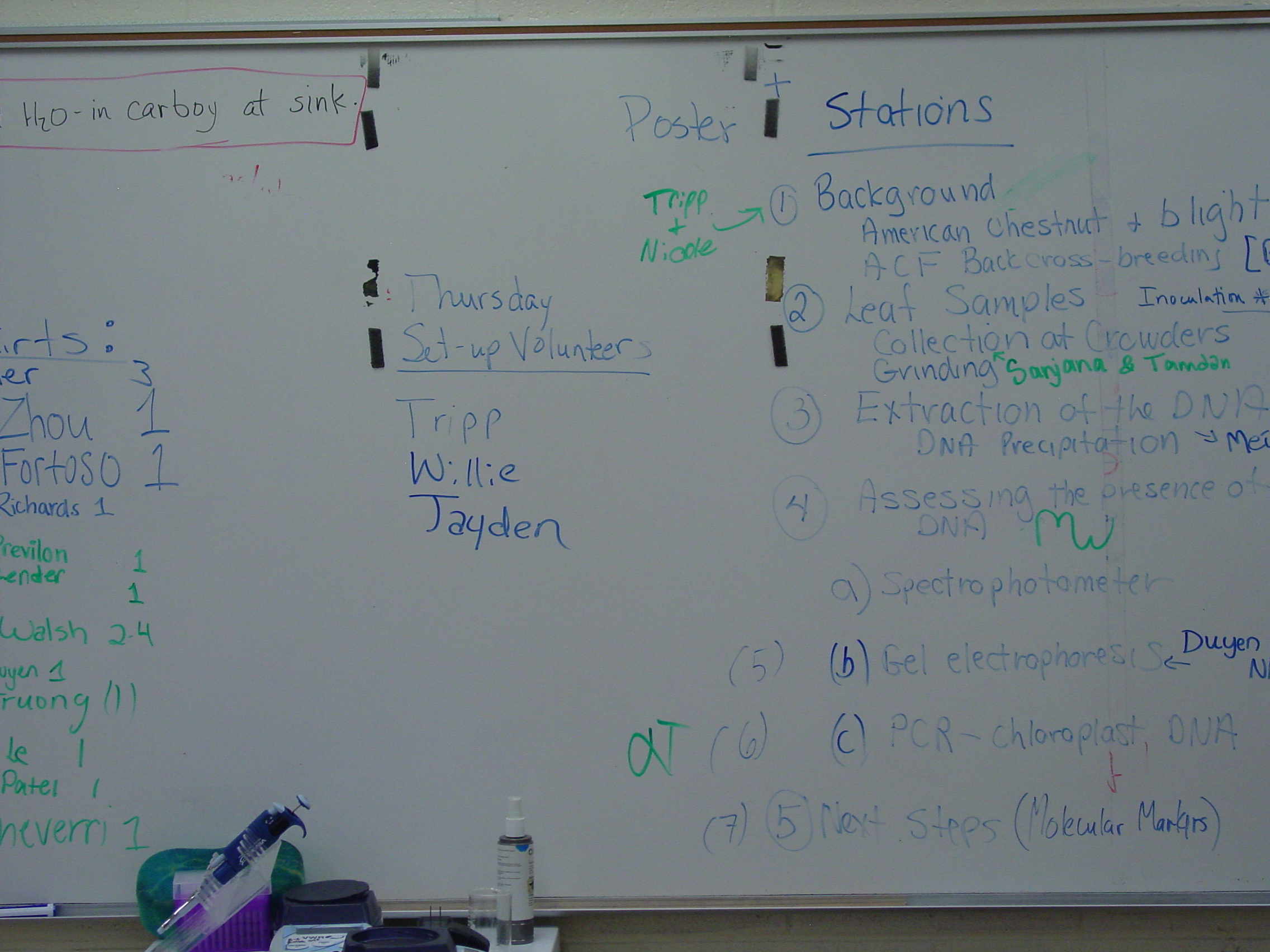
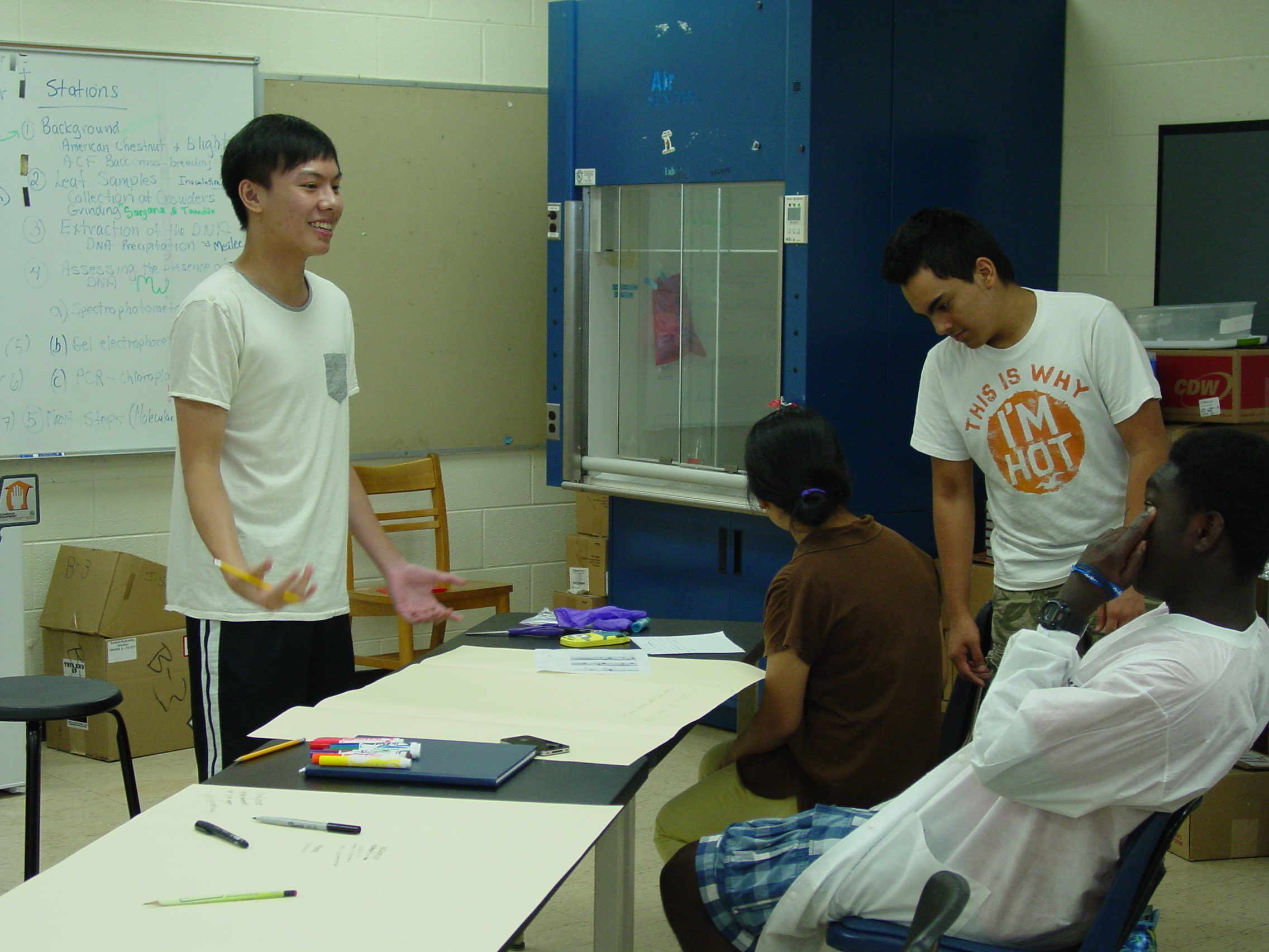
We ran the last gel on our last PCR reactions, and then planned our posters, which we will make tomorrow.
Setting up the Electrophoresis apparatus Pouring buffer in the tank. Loading wells again! Keeping track of the sample loading order DNA bands glowing in UV light Assigning presentation tasks on white board Poster Design sessions. Wednesday, June 25 th, 2014. The B3 Summer Science Camp Blog
This day was our last day of lab work. We prepared our last PCR samples and did our last gel. I think we were all pleased with our gels from
the day before; we all got some DNA and it showed up. Our lab work did not take long, so we spent the rest of time preparing for our scientific poster
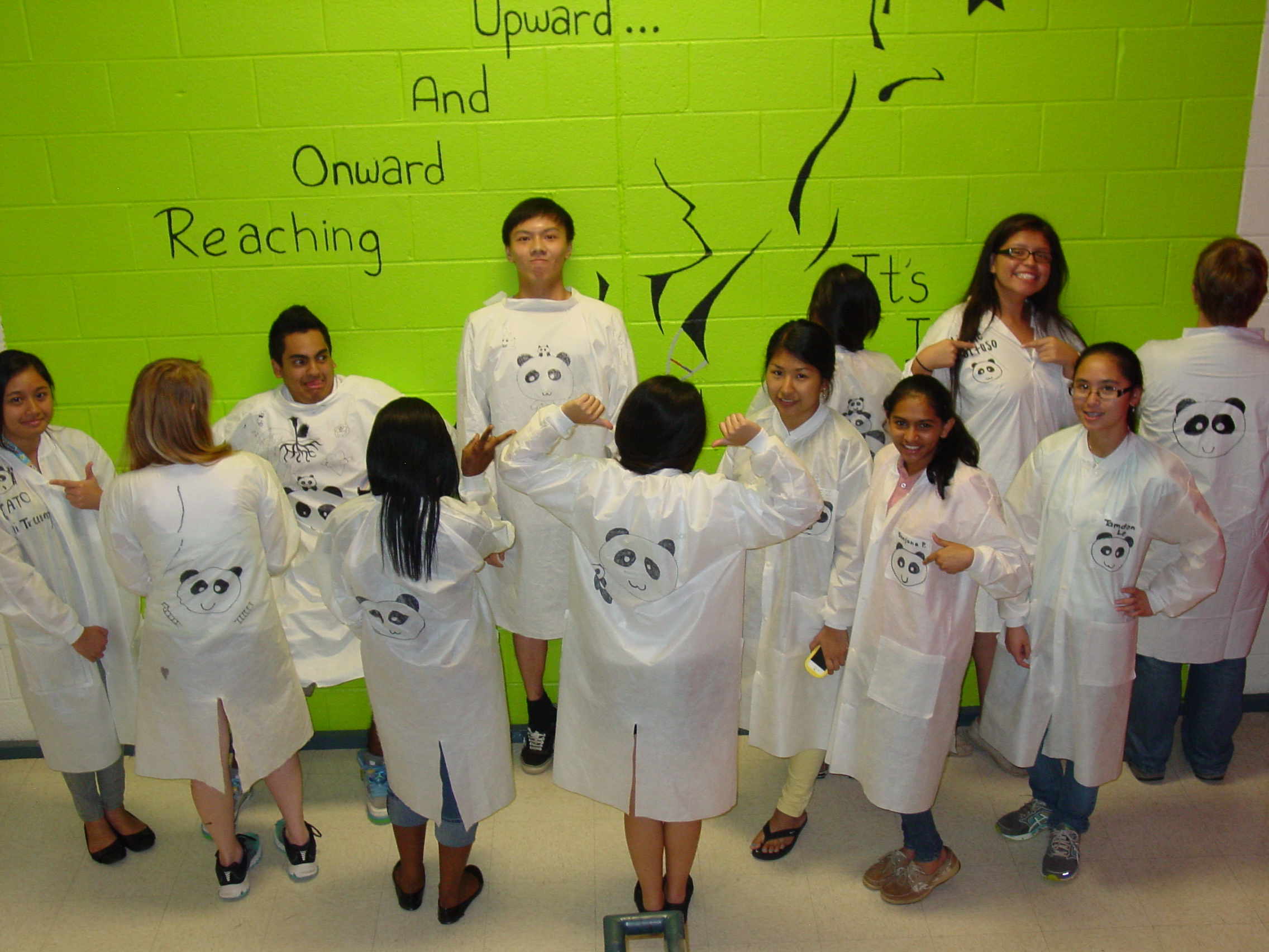
competition, ...or at least the layout. We also took pictures! #pandaclub. We focused on preparing our work for tomorrow to show our friends and families.
It was all work with some play. ----- Maria Echeverri
June 26 th
Cast of Characters
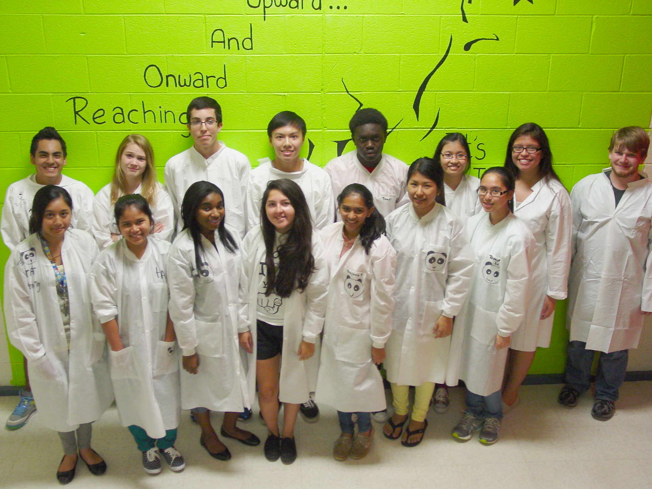
The B3 Class of 2014 is shown below
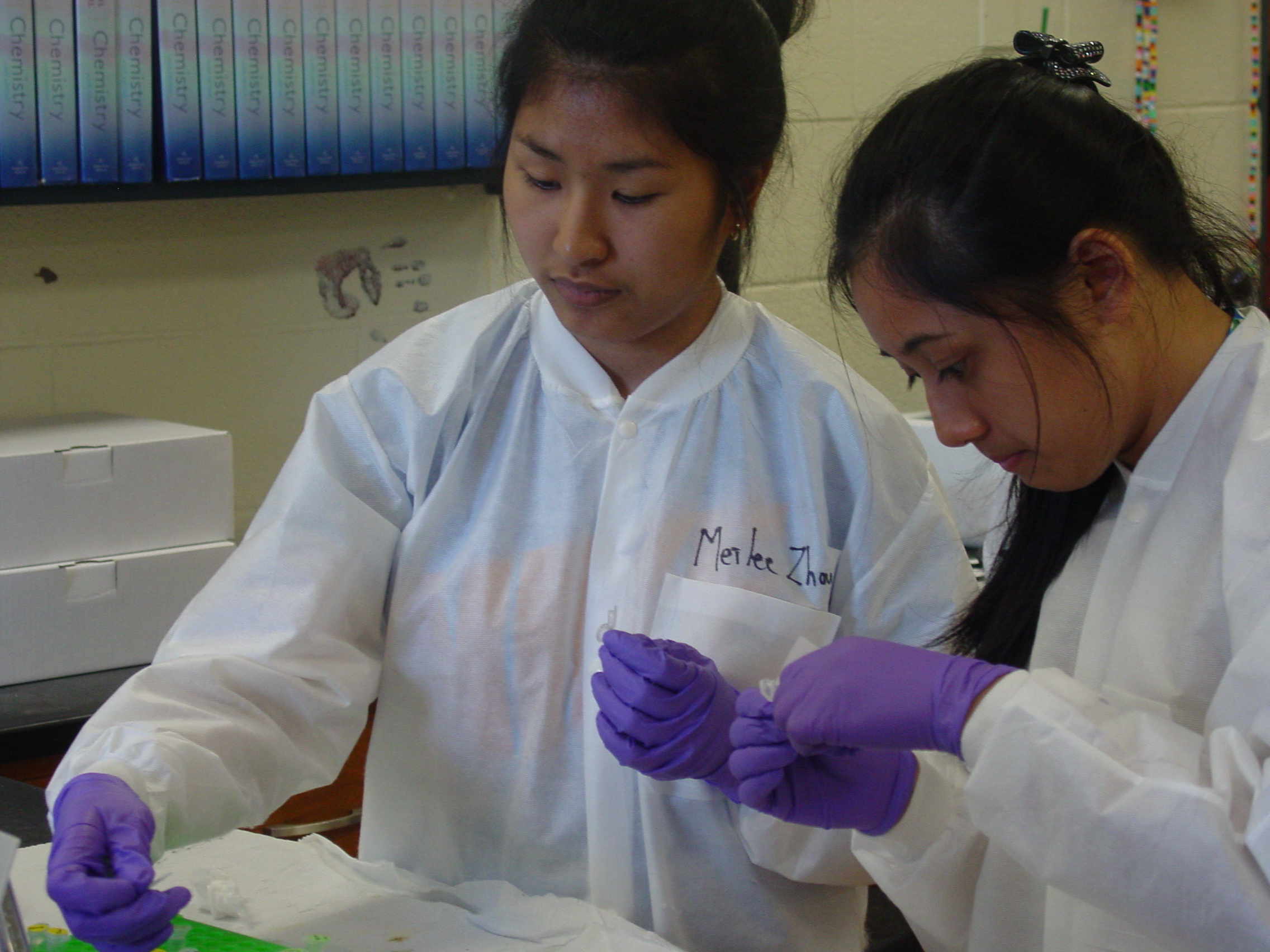
Front Row: Nhi Truong, Hoa Ksor, Doudgie Revilon, Maria Echeverri, Sanjana Patel, Meilee Zhou, Tamden Le
Back Row: Willie Velis, Amara Varney, Tripp Stender, Anthony Ung, James Tweneboah, Duyen Nguyen, Nicole Fortoso, Jayden Walsh (TA)
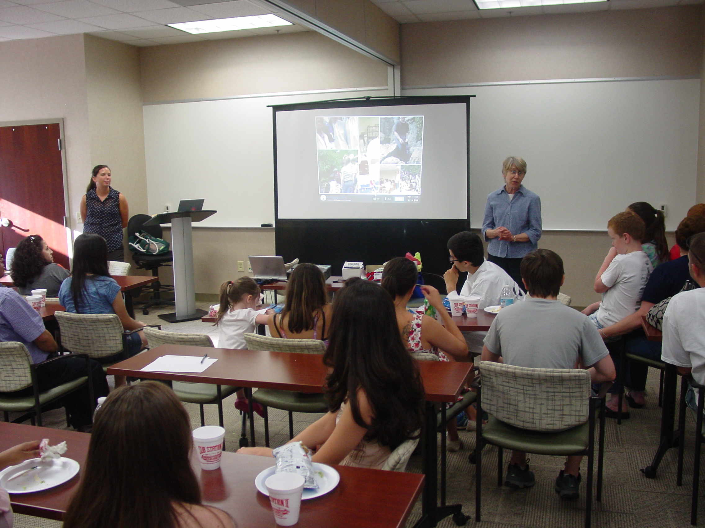
We had the poster and demonstration sessions at the CMC Steele Creek Community Center.
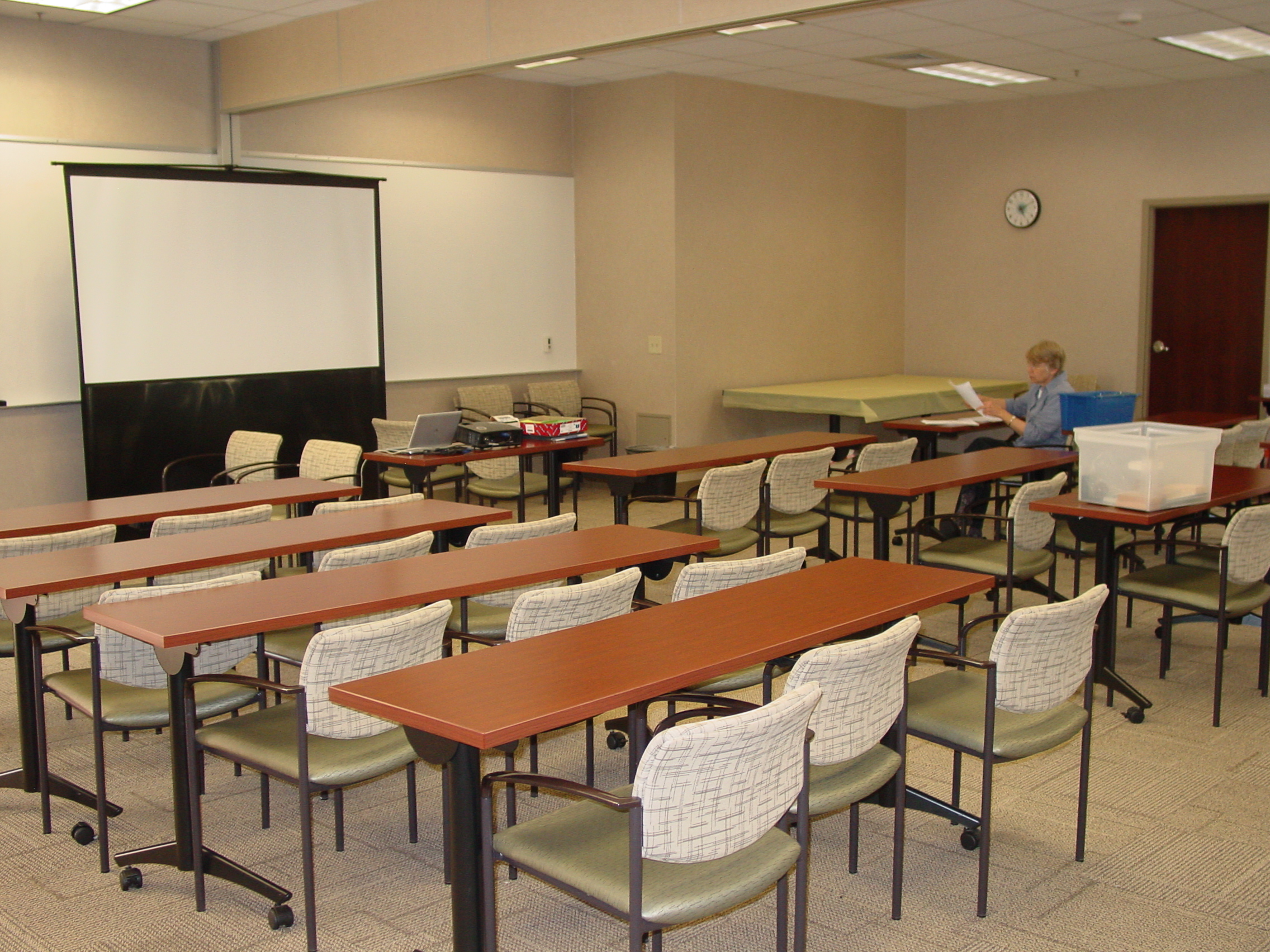
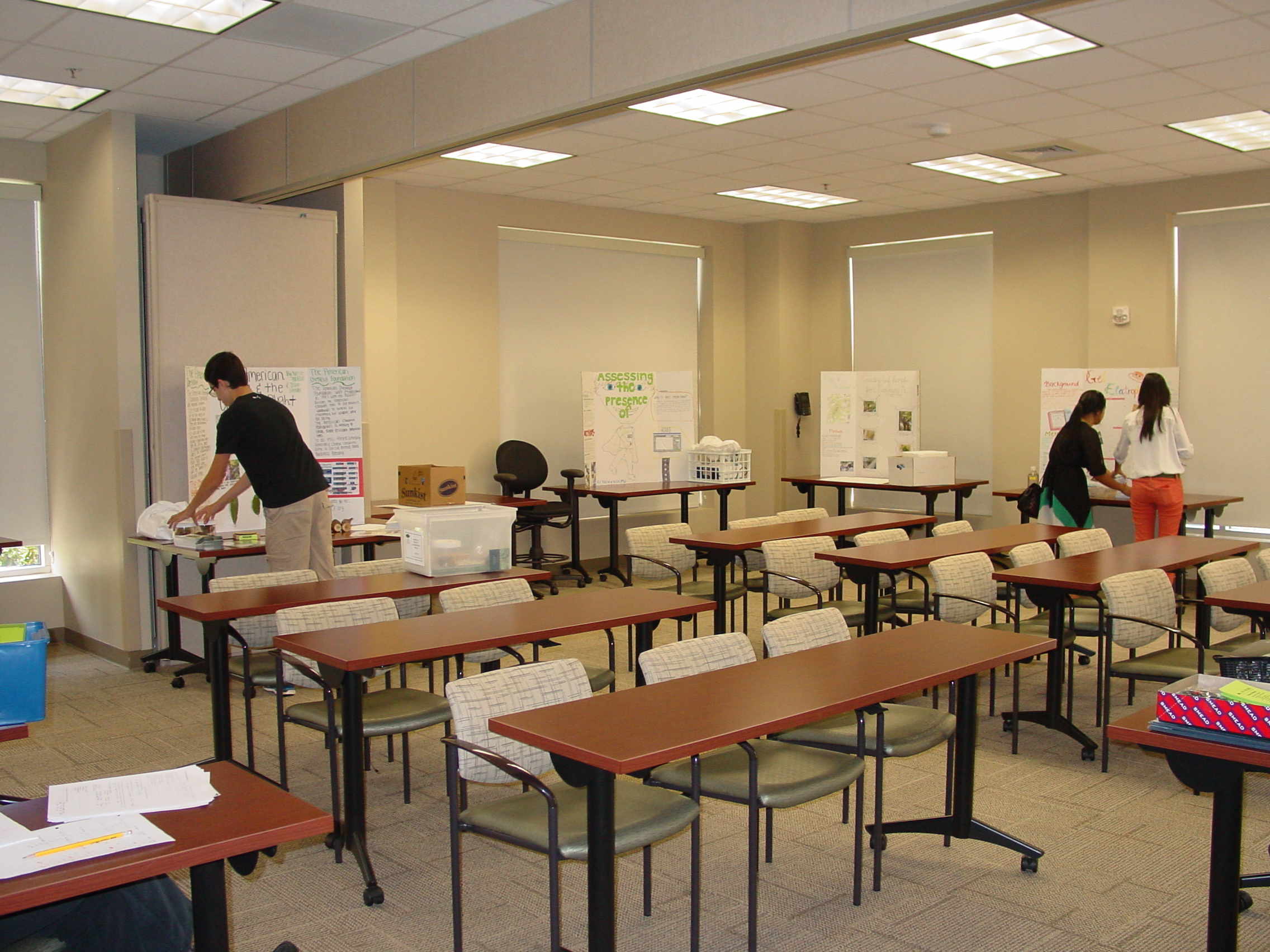
Getting the room and posters set up, and giving a general introduction to guests to guests
Poster and presenters (where we captured them in situ) are shown below.
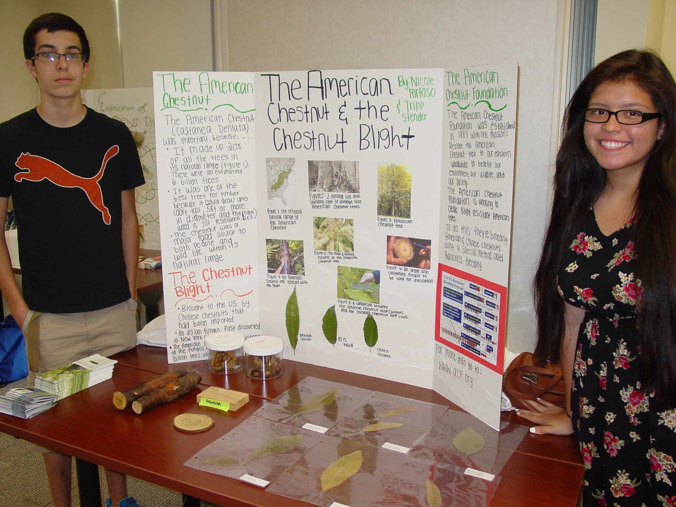
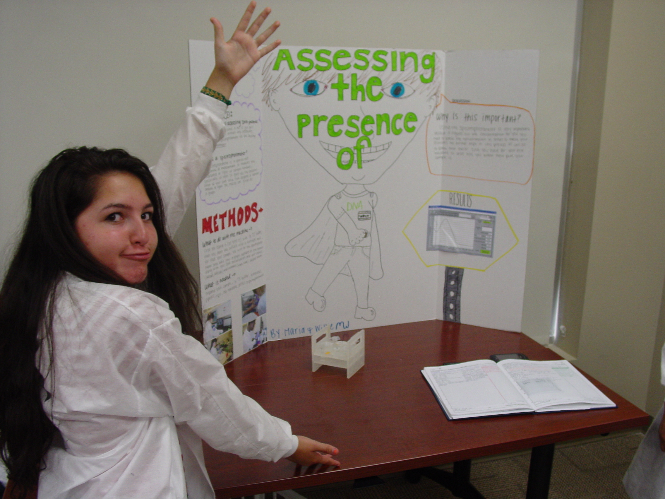
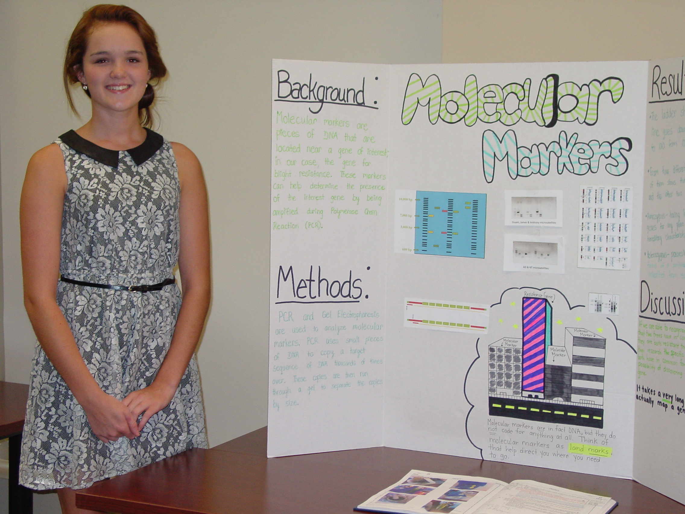
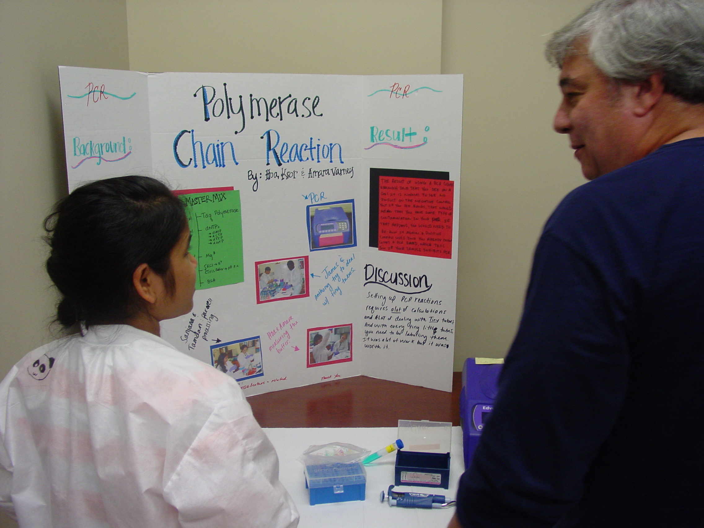
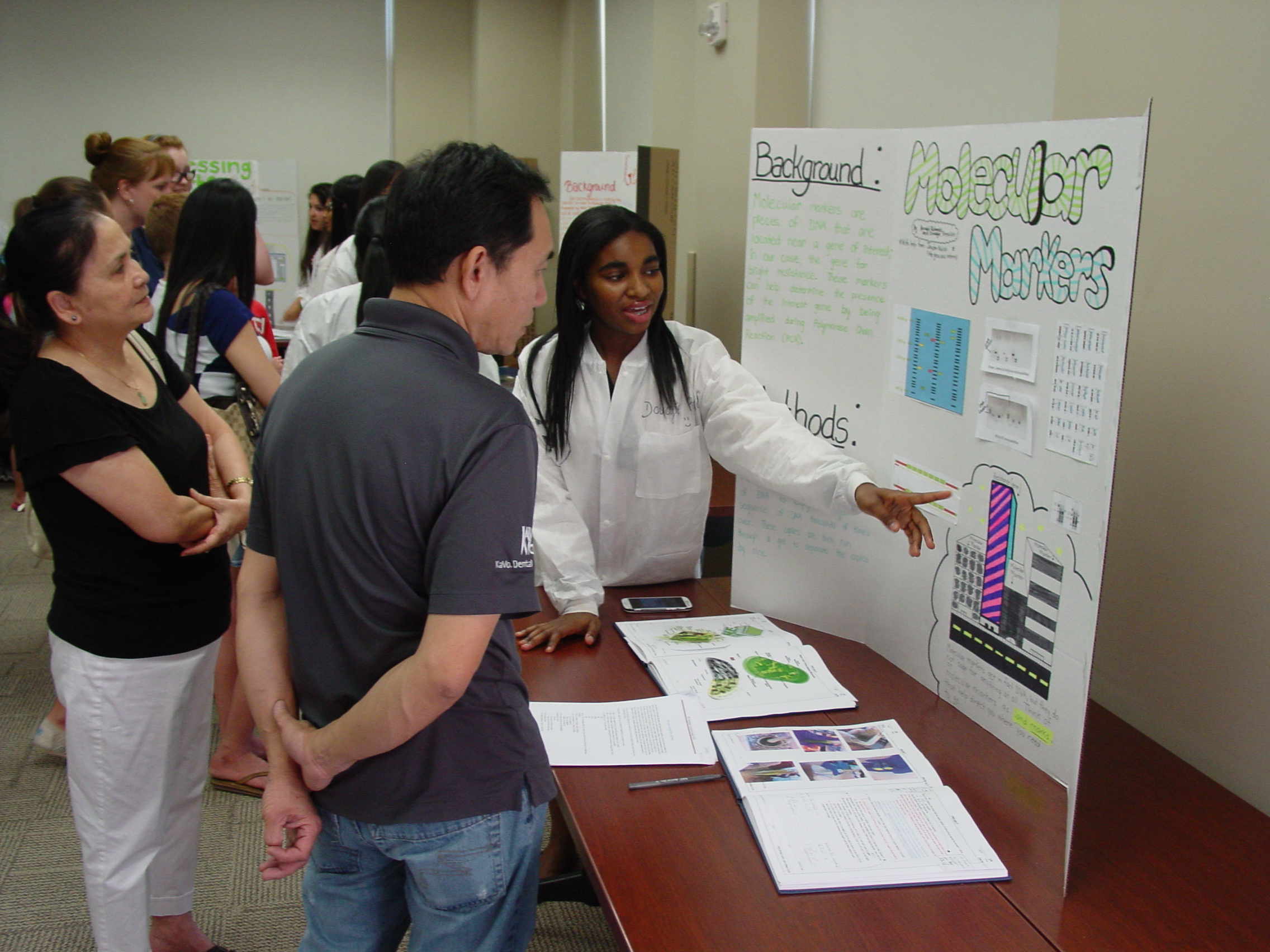
Tripp and Nicole on The American Chestnut and its Blight Sanjana and Tamden on Leaf Processing Anthony and James (not shown) presented the DNA extraction process Hoa and Amara (not shown) presented the PCR method Duyen and Nhi presented the electrophoresis process Maria and Willie (not shown) presented Spectrophotometry Annabel and Doudgie (shown below) presented the Molecular Markers concept
We had a judge (Steve Barilovits) for the posters
Poster judging in process Doudgie explaining the poster Poster winners: Annabel and Doudgie
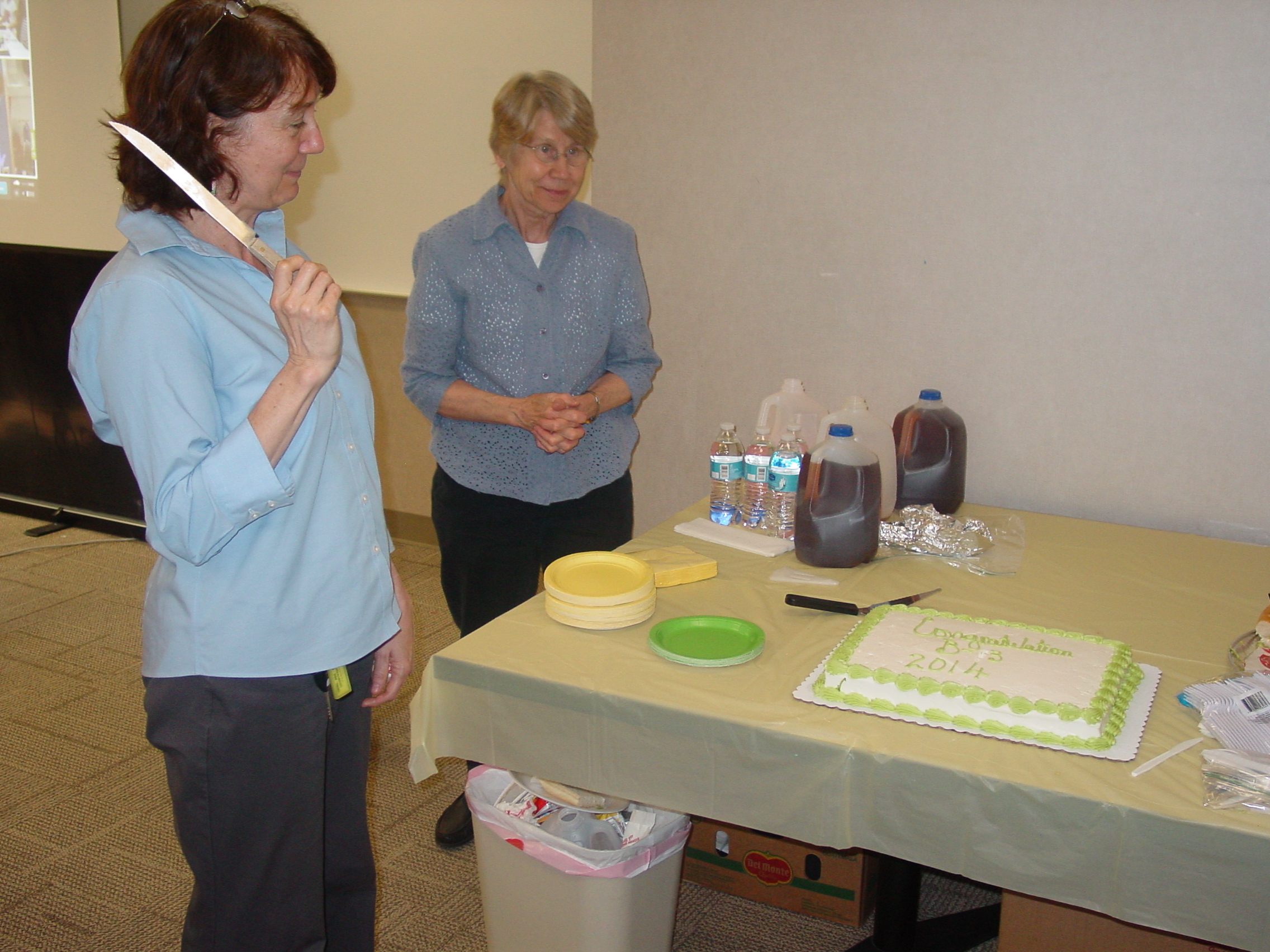
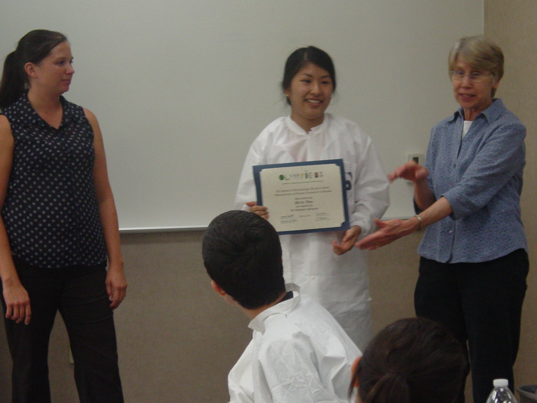
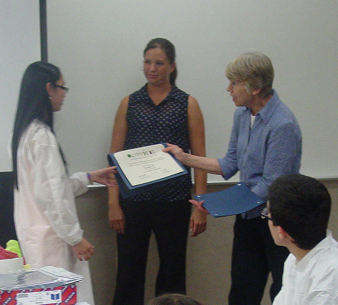
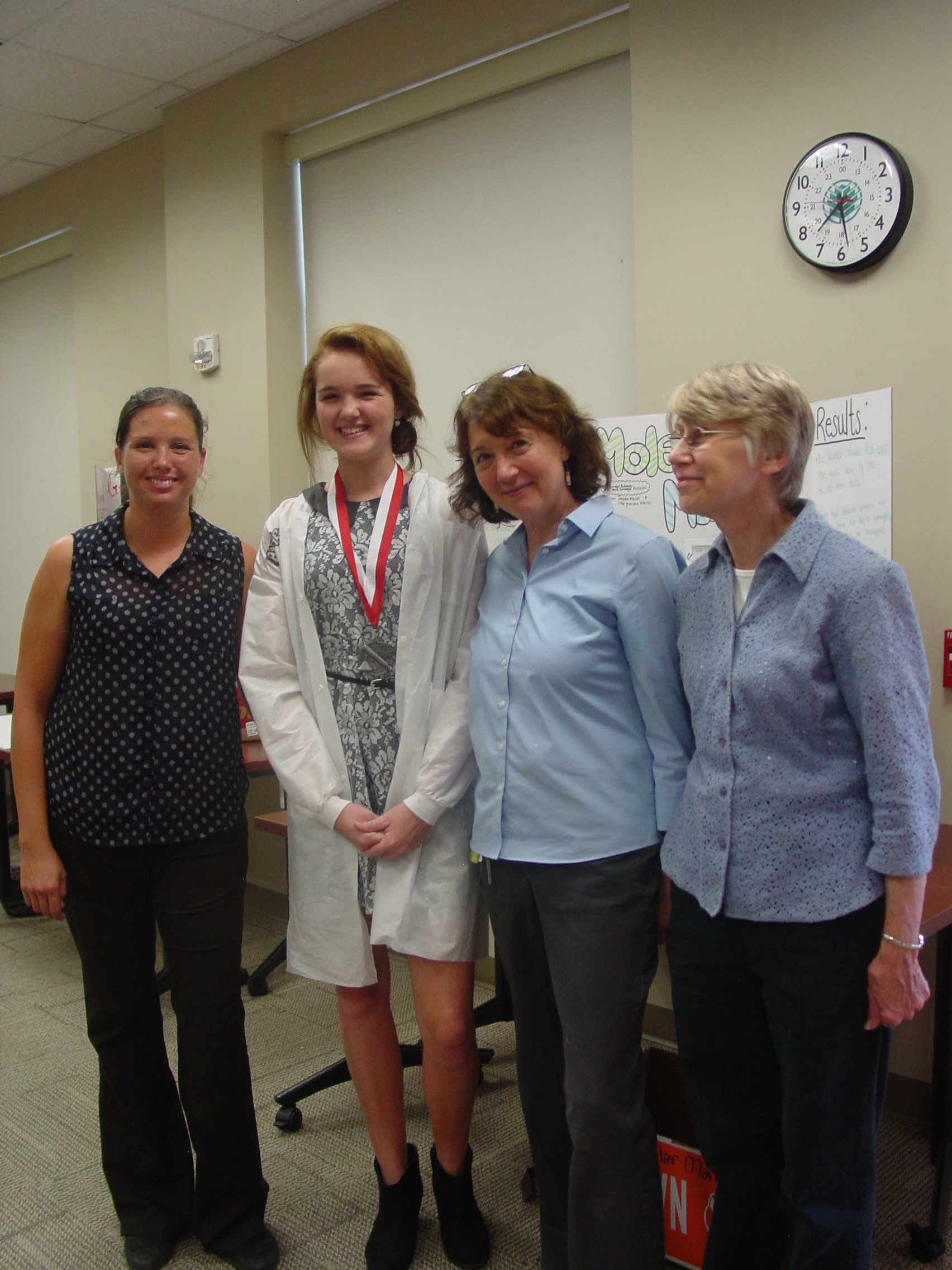
After dinner we had cake while watching awarding of certificates (most people were in motion and we did not get good photos).
Dr. Weller trying to cut identical pieces Meilee is certified Tamden accepts her certificate
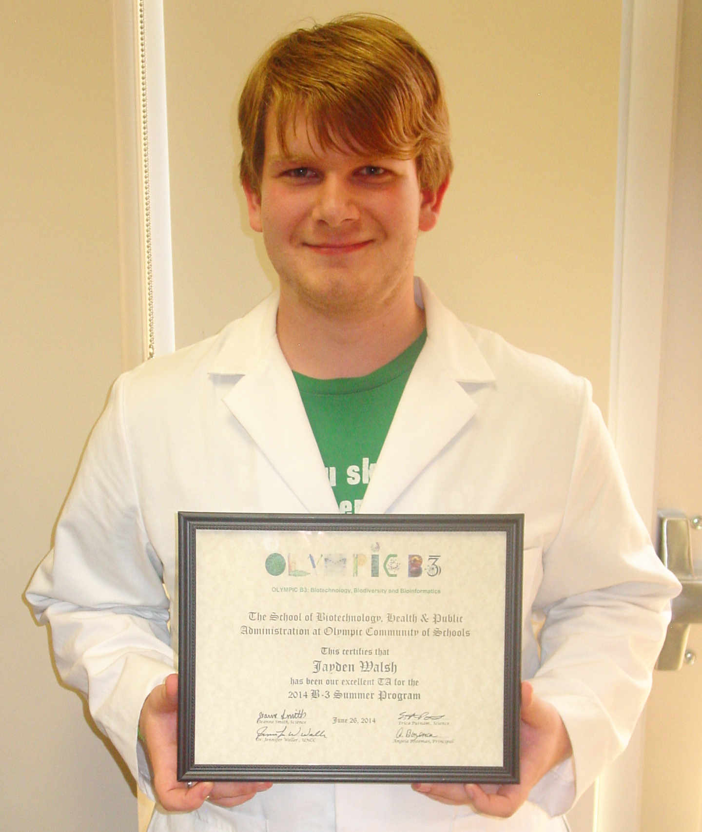
The exceptional efforts of other individuals to the success of the camp were noted as well.
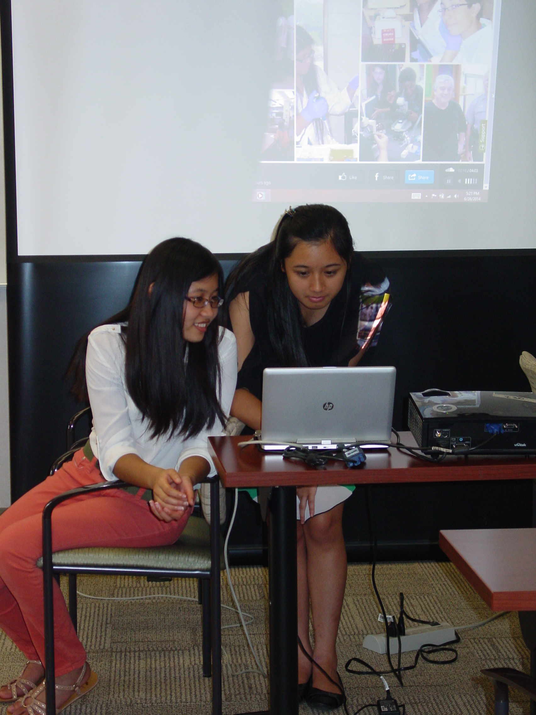
Our TA, Jayden Walsh Our journalist, Annabel Richards Slide show editors, Duyen, Nhi and Maria
June 27 th
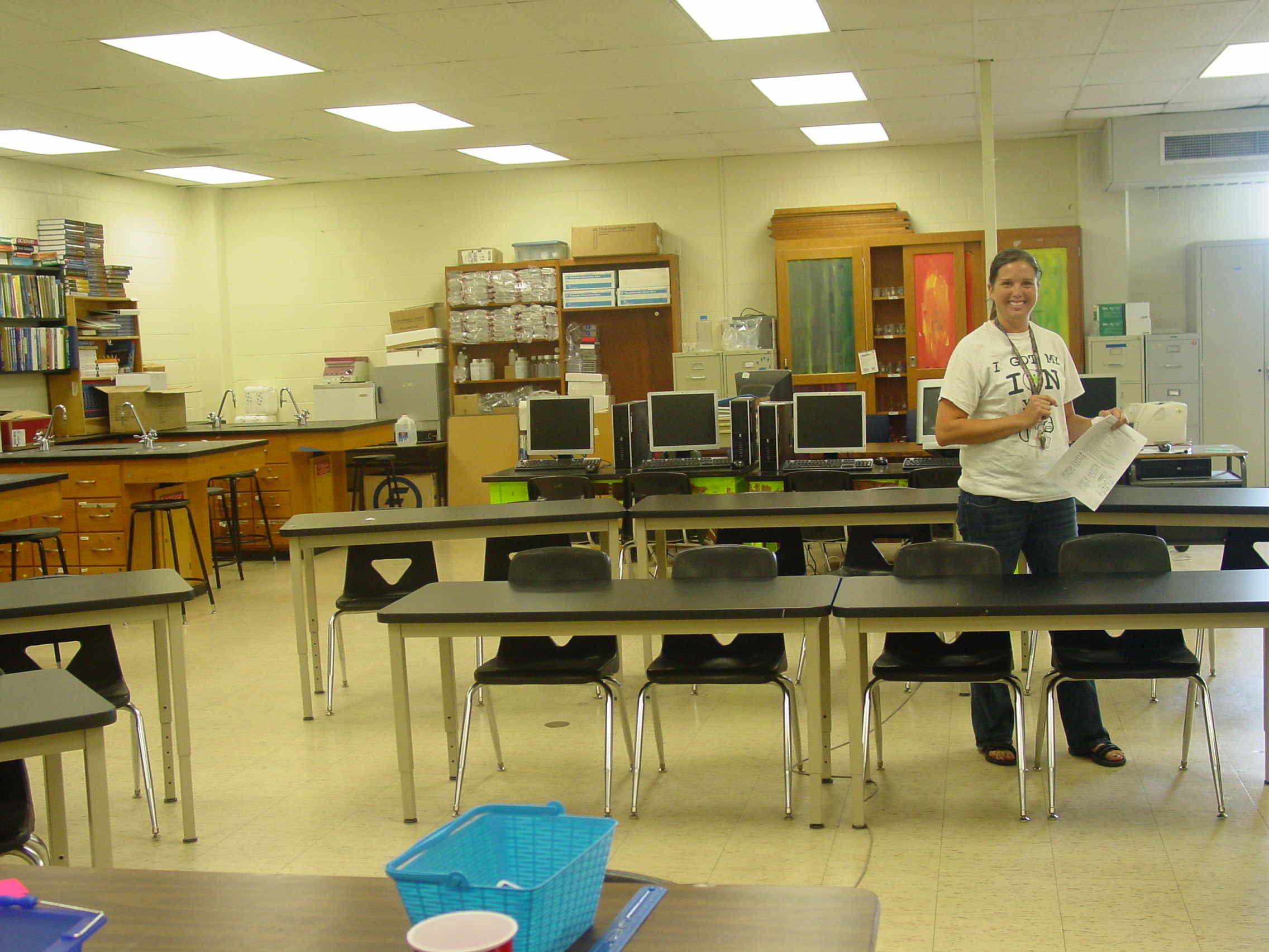
Lab Cleanup Take only pictures, leave only footprints
The lab has to be left in a pristine state, so the instructors spent part of Friday cleaning up.
Thanks to everyone for another successful B3 Camp!
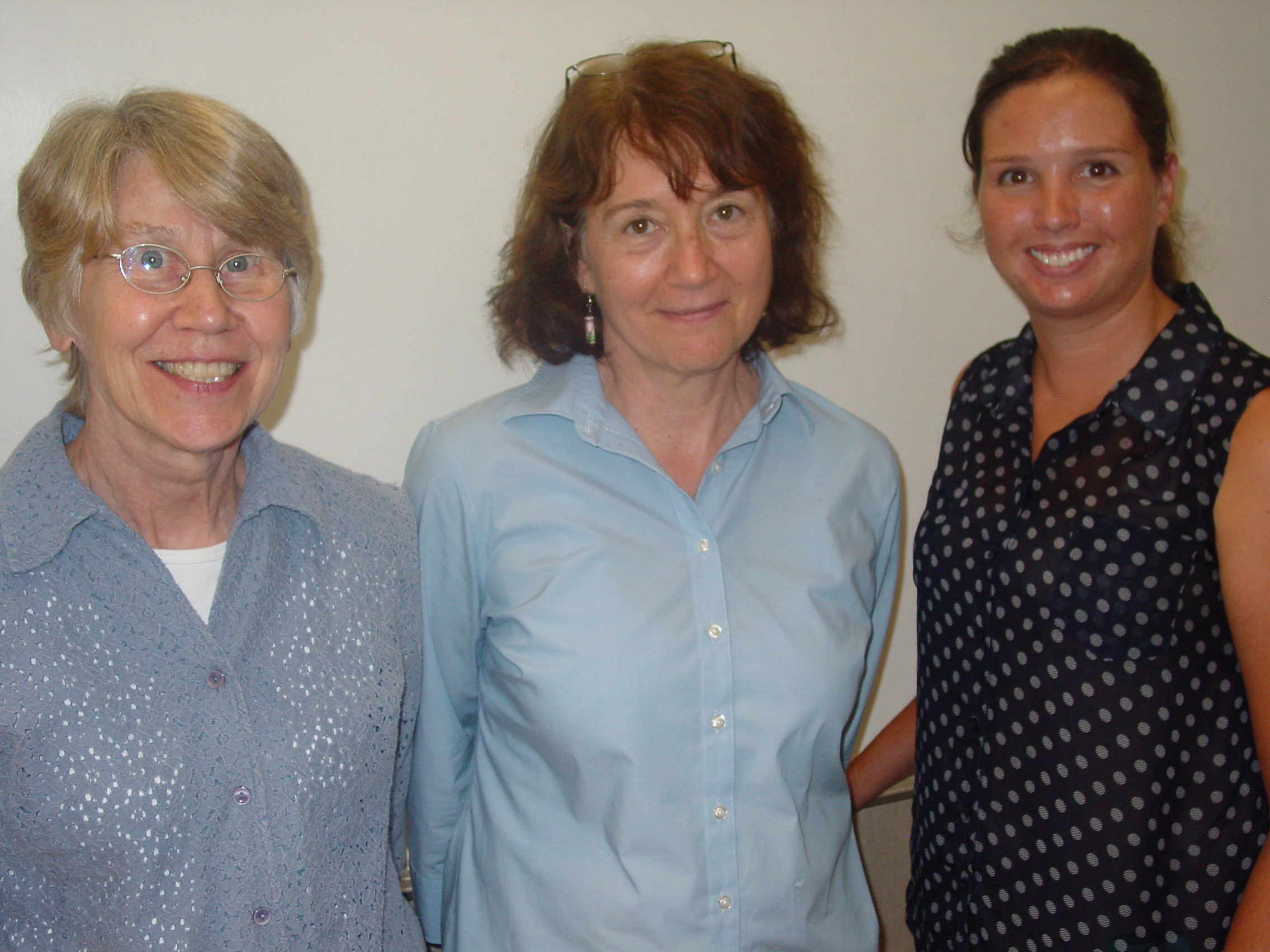
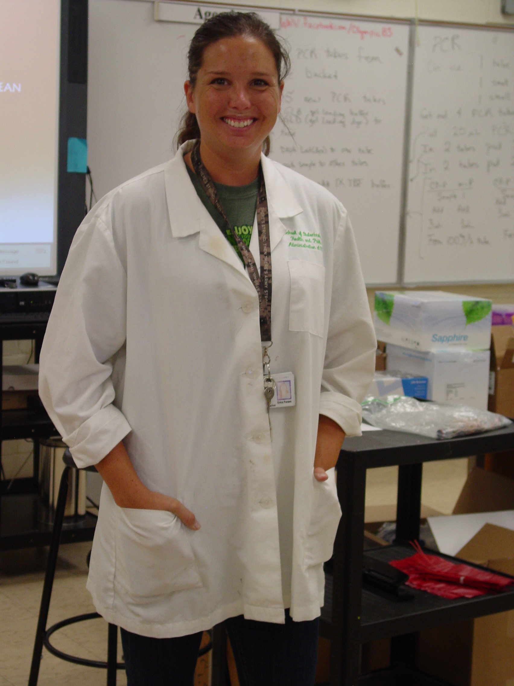
Jeanne, Jennifer and Erica
&
Other Content
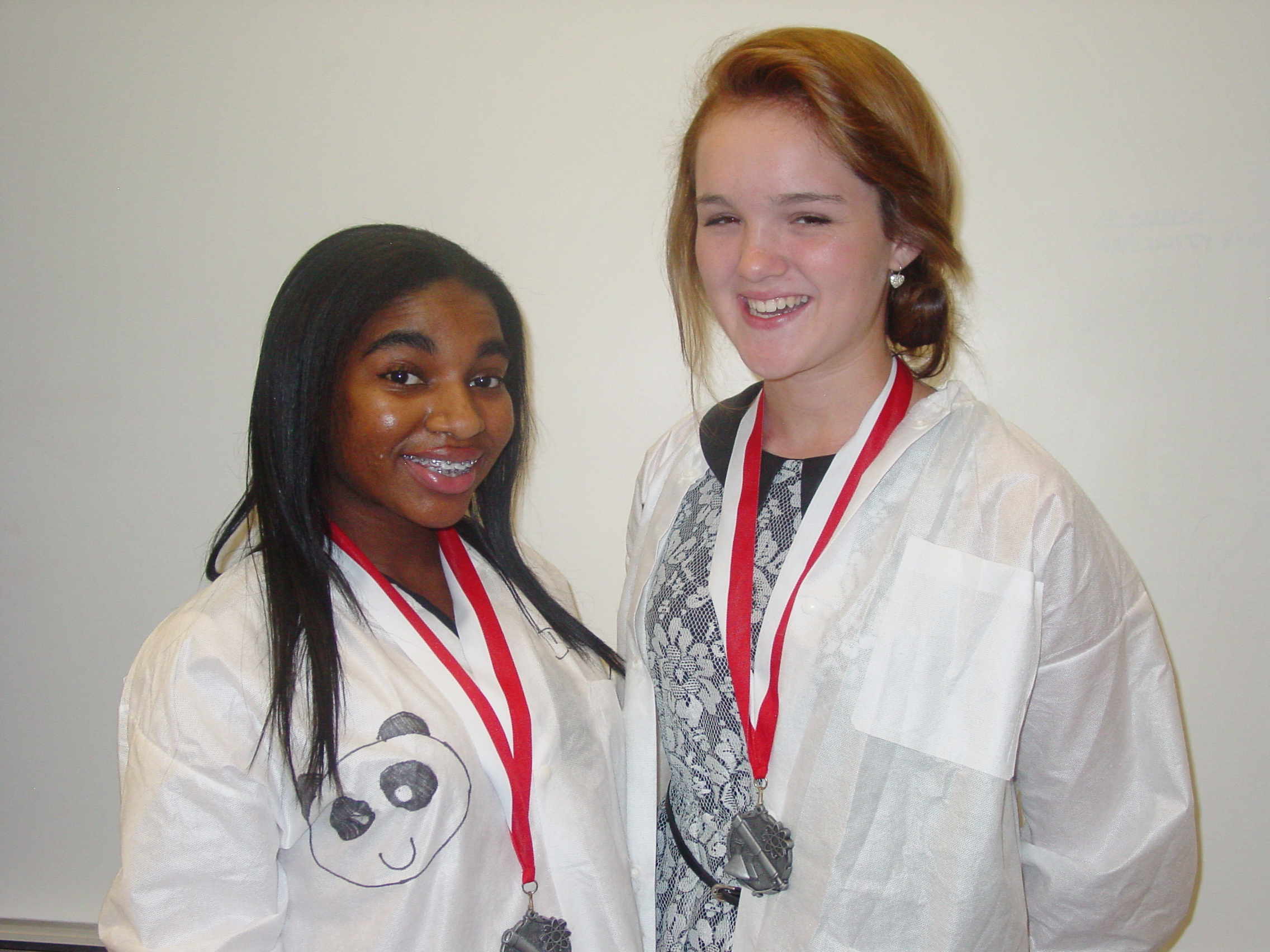
The Panda (Labcoat) Club wishes to make the following statement.
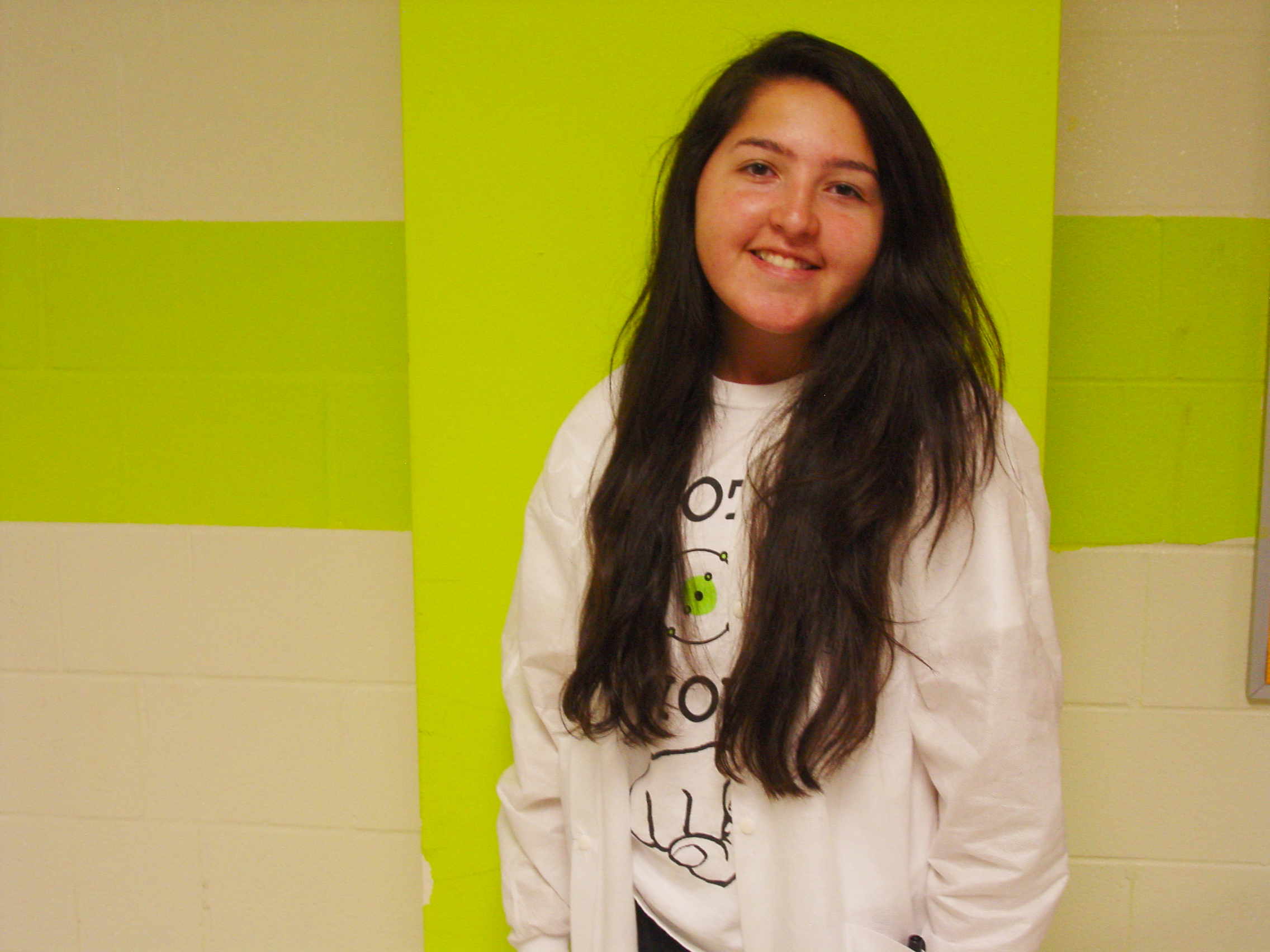
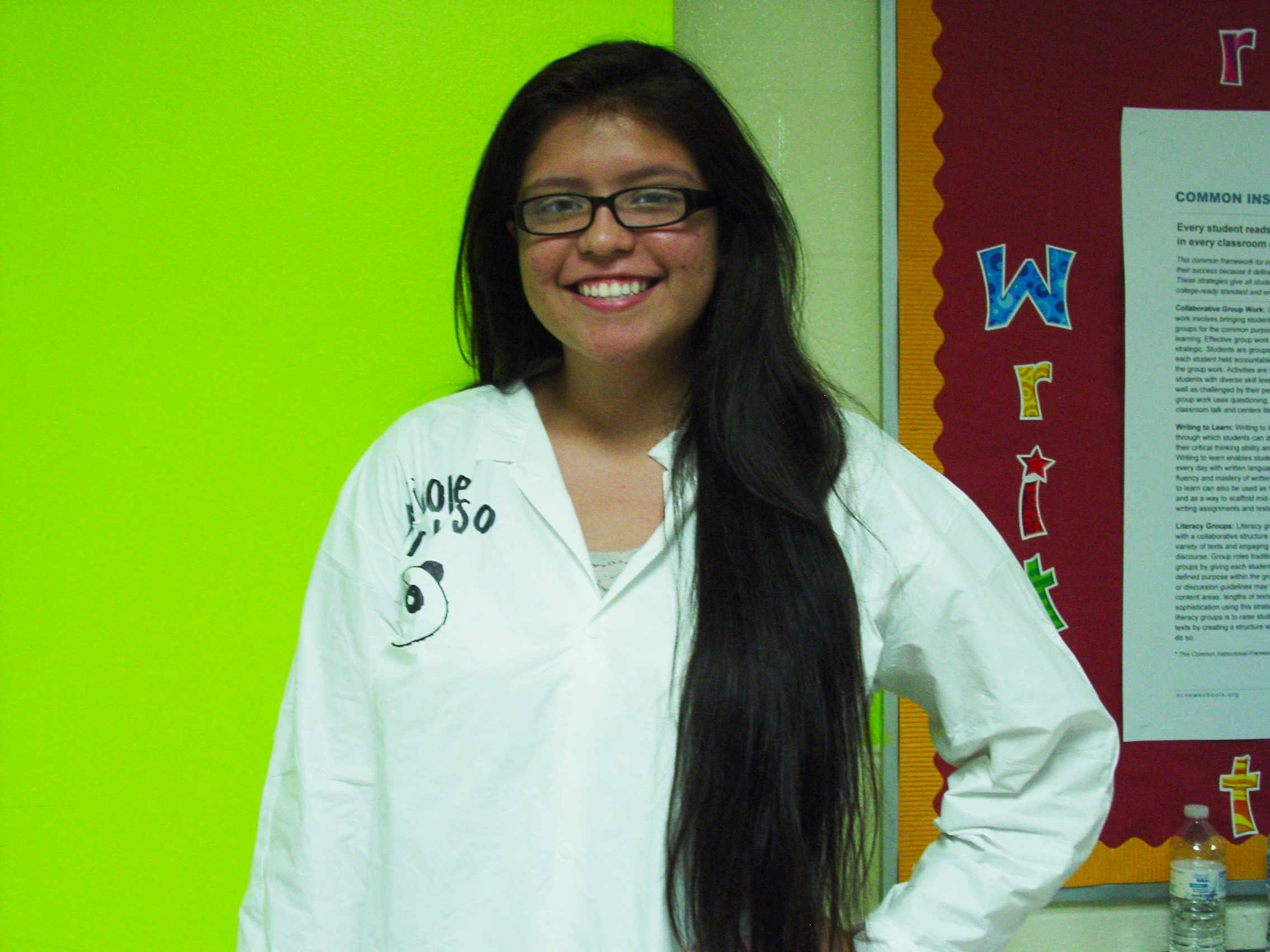
By Request, some individual mug shots.
Maria Nicole Hoa Tamden Duyen Sanjana Doudgie Nhi Meilee Jayden Ms Putnam Ms Smith